J Cardiovasc Ultrasound.
2014 Mar;22(1):43-45. 10.4250/jcu.2014.22.1.43.
Unique Congenital Malformation of the Mitral Valve Associated with Anomalous Coronary Arteries and Stroke
- Affiliations
-
- 1Department of Cardiac Sciences, Libin Cardiovascular Institute, Foothills Medical Center, University of Calgary, Calgary, Alberta, Canada. tarekdeema@hotmail.com
- KMID: 2177457
- DOI: http://doi.org/10.4250/jcu.2014.22.1.43
Abstract
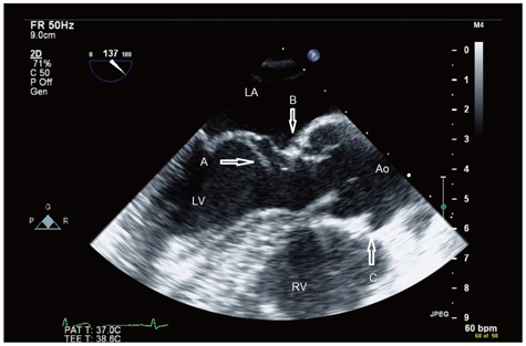
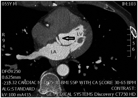
- A 55-year-old male presented with stroke. Transesophageal echocardiogram and cardiac computed tomography revealed an unrecognized congenital malformation of the anterior mitral leaflet associated with anomalous left coronary circumflex artery, arising from the right coronary artery, diagnosed first by echocardiogram. This case represents a unique unforeseen mitral valve anomaly that might be considered as potential cardiac source of embolism. This finding broadens the spectrum of known mitral valve anomalies.
Keyword
MeSH Terms
Figure
Reference
-
1. Séguéla PE, Houyel L, Acar P. Congenital malformations of the mitral valve. Arch Cardiovasc Dis. 2011; 104:465–479.
Article2. Banerjee A, Kohl T, Silverman NH. Echocardiographic evaluation of congenital mitral valve anomalies in children. Am J Cardiol. 1995; 76:1284–1291.
Article3. Bär H, Siegmund A, Wolf D, Hardt S, Katus HA, Mereles D. Prevalence of asymptomatic mitral valve malformations. Clin Res Cardiol. 2009; 98:305–309.
Article4. Park MH, Jung SY, Youn HJ, Jin JY, Lee JH, Jung HO. Blood cyst of subvalvular apparatus of the mitral valve in an adult. J Cardiovasc Ultrasound. 2012; 20:146–149.
Article5. Prifti E, Frati G, Bonacchi M, Vanini V, Chauvaud S. Accessory mitral valve tissue causing left ventricular outflow tract obstruction: case reports and literature review. J Heart Valve Dis. 2001; 10:774–778.6. Cil H, Atilgan ZA, Islamoglu Y, Yavuz C, Tekbas EÖ. Asymptomatic and isolated accessory mitral valve tissue in adult population: three case reports and review of the literature. Eur Rev Med Pharmacol Sci. 2012; 16:Suppl 4. 74–77.7. Singh B, Srinivasa KH, Surangi MJ, Rangan K, Manjunath CN. Anomalous mitral arcade variant with accessory mitral leaflet and chordae presenting for the first time with acute decompensated heart failure in an adult. Echocardiography. 2013; 30:E202–E205.
Article8. Uysal OK, Duran M, Ozkan B, Tekin K, Elbasan Z. Asymptomatic accessory mitral valve tissue diagnosed by echocardiography. Korean Circ J. 2012; 42:800.
Article9. Cohen MS, Herlong RJ, Silverman NH. Echocardiographic imaging of anomalous origin of the coronary arteries. Cardiol Young. 2010; 20:Suppl 3. 26–34.
Article
- Full Text Links
- Actions
-
Cited
- CITED
-
- Close
- Share
- Similar articles
-
- Mitral valve prolapse
- A Case of Isolated Congenital Double-Orifice Mitral Valve
- Congenital Double-Orifice Mitral Valve with Mitral Valve Prolapse and Severe Mitral Regurgitation
- Anomalous Origin of the Left Coronary Artery from the Pulmonary Artery in an Adult: A case report
- Rare Case of Unileaflet Mitral Valve


