Porcelain Heart: Rapid Progression of Cardiac Calcification in a Patient with Hemodialysis
- Affiliations
-
- 1Division of Cardiology, Department of Internal Medicine, Pohang St. Mary's Hospital, Pohang, Korea.
- 2Division of Cardiology, Department of Internal Medicine, Seoul St. Mary's Hospital, School of Medicine, The Catholic University of Korea, Seoul, Korea. younhj@catholic.ac.kr
- 3Department of Radiology, Pohang St. Mary's Hospital, Pohang, Korea.
- 4Division of Cardiology, Department of Internal Medicine, Uijeongbu St. Mary's Hospital, School of Medicine, The Catholic University of Korea, Uijeongbu, Korea.
- KMID: 2177407
- DOI: http://doi.org/10.4250/jcu.2012.20.4.193
Abstract
- Cardiac calcification usually occurs in patients with end-stage renal disease. However, rapid progression of cardiac calcification is rarely associated with secondary hyperparathyroidism of end-stage renal disease. We report a patient with end-stage renal disease who showed moderate left ventricular hypertrophy at the first echocardiography, and showed severe myocardial calcification and severe mitral valve stenosis 4 years later. We suspected a rapid progression 'porcelain heart' cardiomyopathy secondary to hyperparathyroidism of end-stage renal disease. The patient underwent parathyroidectomy, and considered mitral valve replacement.
Keyword
MeSH Terms
Figure
-
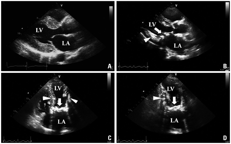
Fig. 1 Changes of transthoracic echocardiography. The echocardiogram on parasternal long axis view shows moderate LVH in 2007 (A). Follow up echocardiogram shows extensive myocardial calcification (arrowhead) and severe mitral stenosis with a mitral valve calcification (arrow) in 2011 (B, C and D). LVH: left ventricular hypertrophy, LV: left ventricle, LA: left atrium.
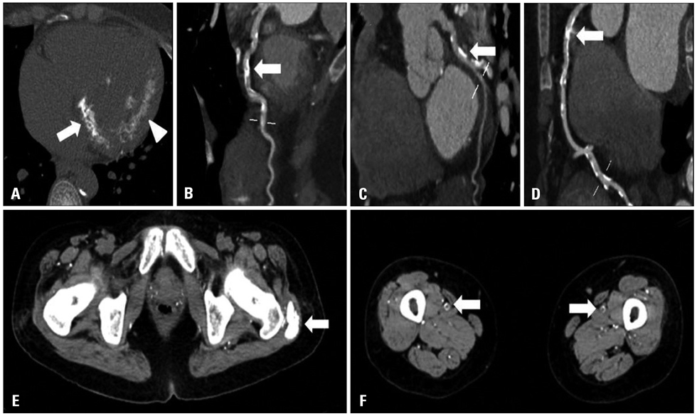
Fig. 2 Cardiac CT (A-D) and peripheral CT (E and F) shows extensive calcification. Cardiac CT shows severe mitral valve calcification (arrow) and myocardial calcification (arrowhead, A), diffuse calcification of LAD coronary artery (B), LCX coronary artery (C) and RCA coronary artery (D). Non enhanced CT shows muscular calcification (arrow, E) and superficial femoral artery calcification (arrows, F). LAD: left anterior descending, LCX: left circumflex, RCA: right coronary artery.
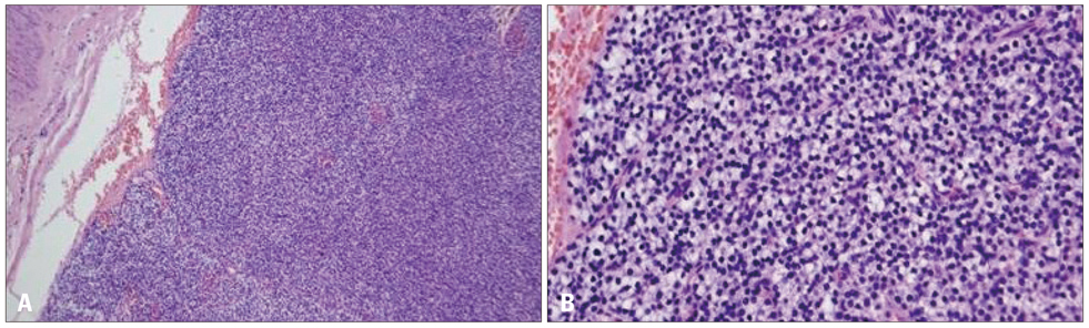
Fig. 3 Microscopic findings. Histology exam shows parathyroid tissue with diffuse hyperplasia of chief cells (A: H&E stain, × 100; B: H&E stain, × 200).
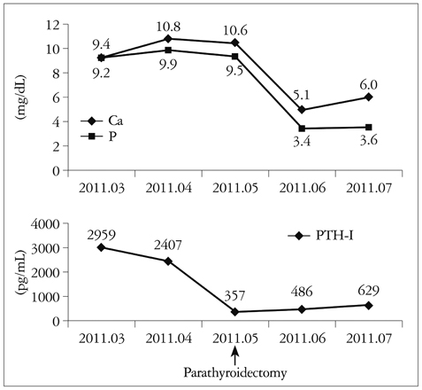
Fig. 4 Change of Ca, P and PTH-I level after parathyroidectomy. PTH-I: immuno-reactive parathyroid hormone.
Reference
-
1. Raggi P, Boulay A, Chasan-Taber S, Amin N, Dillon M, Burke SK, Chertow GM. Cardiac calcification in adult hemodialysis patients. A link between end-stage renal disease and cardiovascular disease? J Am Coll Cardiol. 2002. 39:695–701.
Article2. Freeman J, Dodd JD, Ridge CA, O'Neill A, McCreery C, Quinn M. "Porcelain heart" cardiomyopathy secondary to hyperparathyroidism: radiographic, echocardiographic, and cardiac CT appearances. J Cardiovasc Comput Tomogr. 2010. 4:402–404.
Article3. Aras D, Topaloglu S, Demirkan B, Deveci B, Ozeke O, Korkmaz S. Porcelain heart: a case of massive myocardial calcification. Int J Cardiovasc Imaging. 2006. 22:123–126.
Article4. Kempf AE, Momeni MG, Saremi F. Myocardial calcinosis in chronic renal failure. J Radiol Case Rep. 2009. 3:16–19.5. London GM, Guérin AP, Marchais SJ, Métivier F, Pannier B, Adda H. Arterial media calcification in end-stage renal disease: impact on all-cause and cardiovascular mortality. Nephrol Dial Transplant. 2003. 18:1731–1740.
Article6. Fraser WD. Hyperparathyroidism. Lancet. 2009. 374:145–158.
Article7. Wang AY, Ho SS, Wang M, Liu EK, Ho S, Li PK, Lui SF, Sanderson JE. Cardiac valvular calcification as a marker of atherosclerosis and arterial calcification in end-stage renal disease. Arch Intern Med. 2005. 165:327–332.
Article8. Kuzela DC, Huffer WE, Conger JD, Winter SD, Hammond WS. Soft tissue calcification in chronic dialysis patients. Am J Pathol. 1977. 86:403–424.9. Isotalo PA, Halil A, Green M, Tang A, Lach B, Veinot JP. Metastatic calcification of the cardiac conduction system with heart block: an under-reported entity in chronic renal failure patients. J Forensic Sci. 2000. 45:1335–1338.
Article10. Parfrey PS, Foley RN. The clinical epidemiology of cardiac disease in chronic renal failure. J Am Soc Nephrol. 1999. 10:1606–1615.
Article11. Moe SM, Chen NX. Mechanisms of vascular calcification in chronic kidney disease. J Am Soc Nephrol. 2008. 19:213–216.12. Maher ER, Young G, Smyth-Walsh B, Pugh S, Curtis JR. Aortic and mitral valve calcification in patients with end-stage renal disease. Lancet. 1987. 2:875–877.
Article13. Fujise K, Amerling R, Sherman W. Rapid progression of mitral and aortic stenosis in a patient with secondary hyperparathyroidism. Br Heart J. 1993. 70:282–284.
Article14. Massry SG, Smogorzewski M. Management of vascular calcification in CKD patients. Semin Nephrol. 2006. 26:38–41.
Article
- Full Text Links
-
- Actions
-
Cited
- CITED
-
- Close
- Share
-
- Similar articles
-
- A Case of Recurrent Thrombus Associated with Left Atrial Calcification
- Clinical Implication of Vascular Calcification in Patients Undergoing Hemodialysis: The End or the Beginning of Disease
- Massive Left Atrial Calcification Associated with Mitral Valve Replacement
- Severe Calcification of the Left Atrial Wall with Left Atrial Thrombi and an Axillary Hematoma
- A Case of Metastatic Calcification Occurring at Sternoclavicular Joint in a Patient Receiving Maintenance Hemodialysis
