Assessment of Mitral Valve Complex by Three-Dimensional Echocardiography: Therapeutic Strategy for Functional Mitral Regurgitation
- Affiliations
-
- 1Department of Cardiology, Kawasaki Medical School, Kurashiki, Japan. kyoshida@med.kawasaki-m.ac.jp
- KMID: 2177368
- DOI: http://doi.org/10.4250/jcu.2012.20.2.69
Abstract
- The mitral valve complex is consisted of annulus, leaflets, chordae tendineae, papillary muscle (PMs) and surrounding left ventricle. Functional mitral regurgitation (MR) results from left ventricular remodeling such as dilatation or distortion, which displaces the PMs and then tethers the mitral leaflets, restricting leaflet coaptation. Undersized annuloplasty, which has been widely accepted as a simple and effective procedure for functional MR, sometimes worsens the tethering of posterior leaflet and induces recurrent MR. In order to overcome such problems, several additional procedures to the simple annuloplasty have been produced. Three dimensional echocardiography plays an essential role to understand the geometry of mitral valve complex and contributes greatly to decision making of the surgical strategy in functional MR and its postoperative assessment.
MeSH Terms
Figure
-

Fig. 1 Mitral valve complex. The whole mitral valve complex geometry consisted of annulus, leaflets, chordae, papillary muscles and surrounding left ventricle. Mitral annulus shows a nonplanar saddle-shaped configuration in healthy individual. There are two peaks at the aortic insertion and posterior left ventricular wall and two low troughs closest to the apex located medially and laterally (Otto1) and Levine et al.2) with permission). LA: left atrium, LV: left ventricle.
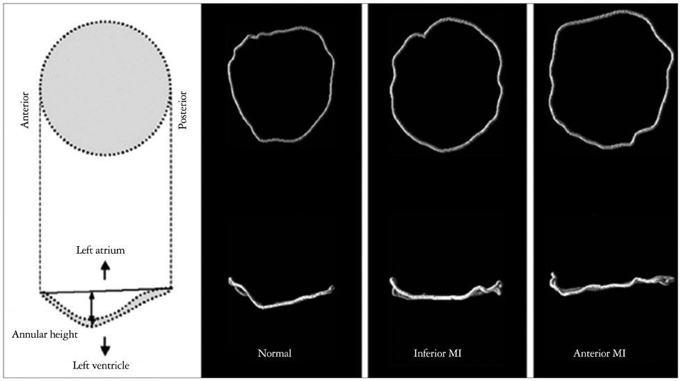
Fig. 2 Mitral annulus configuration. Three dimensional echocardiography allowed us to visualize the dilatation and flattening of the mitral annulus in patients with ischemic mitral regurgitation and more deformation in anterior infarction compared to posterior infarction (Watanabe et al.23) with permission). MI: myocardial infarction.
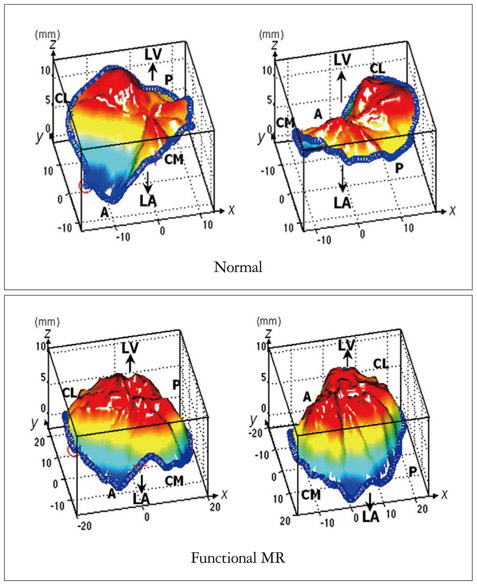
Fig. 3 Visualizing the annular flattening and leaflet tenting. The reconstructed three-dimensional (3D) images by Real View allow us to understand the annular flattening or mitral leaflet tenting in patients with functional mitral regurgitation (MR) and it also can be quantitatively measured and analyzed in 3D spaces, which is impossible to do in two-dimensional images (Watanabe et al.22) with permission).
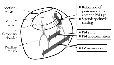
Fig. 4 Surgical procedures for subvalvular apparati or left ventricle. Undersized annuloplasty sometimes fails to ameliorate mitral regurgitation (MR) in patient with severely tethered mitral leaflet and worsen the tethering of posterior leaflet, resulting recurrent MR. In addition, undersized annuloplasty has a possibility to induce functional mitral stenosis. Therefore, additive procedures should be considered for patients with severely tethered valve. PM: papillary muscle.
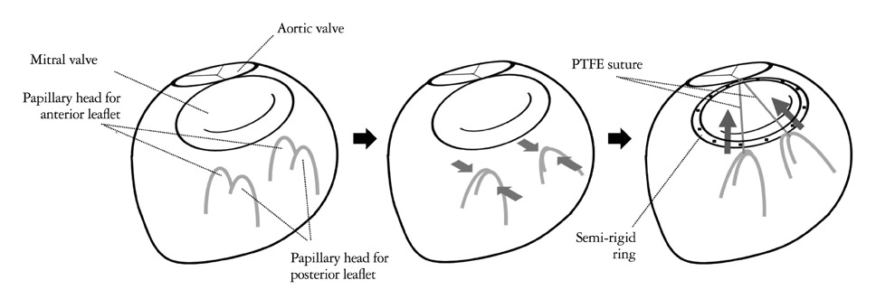
Fig. 5 Papillary heads optimization method. In Papillary heads optimization method, the papillary muscle (PM) head for posterior leaflet was connected to the PM head for anterior leaflet by using a PTFE suture in each PM. After annuloplasty by a semi-rigid ring, its arms were re-suspended towards the mid-anterior mitral annulus and the sutures were passed through it and tied after the length adjustment. PTFE: polytetrafluoroethylene.
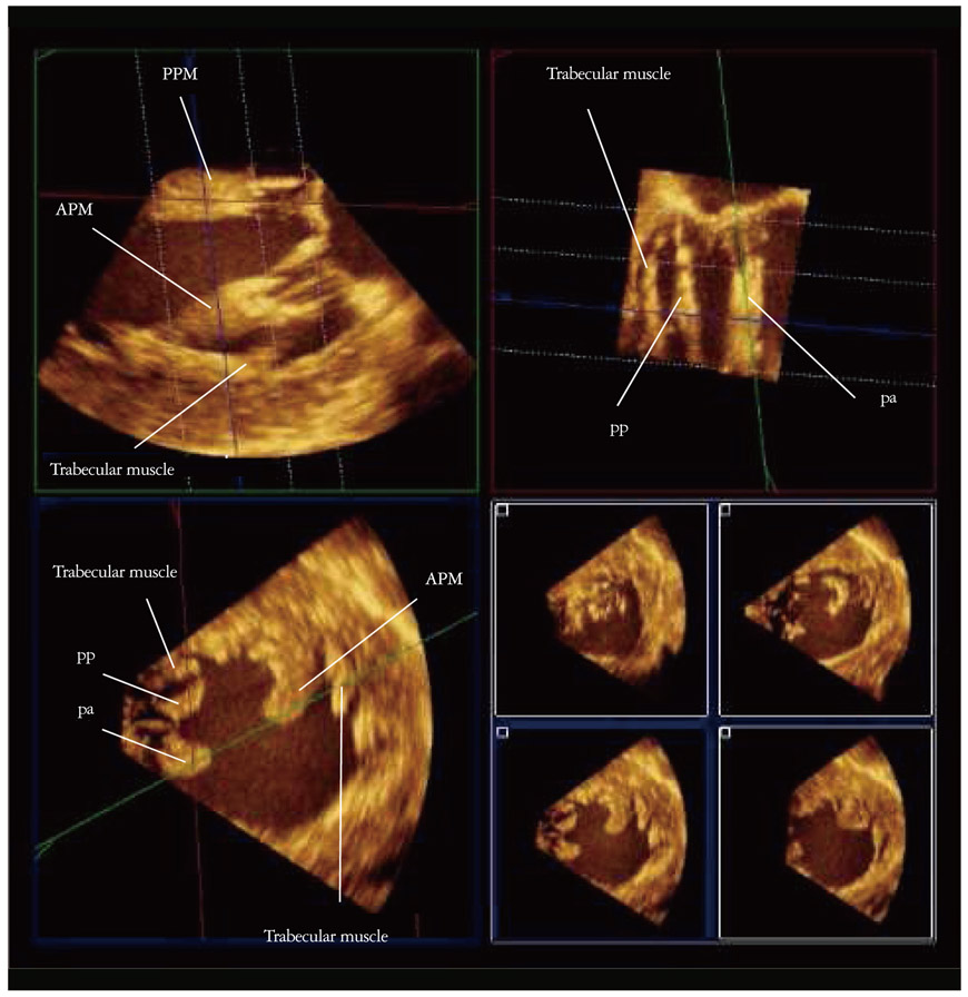
Fig. 6 Visualizing the submitral structure by transgastric approach. Visualizing submitral structure at mid systole from a three-dimensional dataset acquired by transgastric approach using QLAB (Philips Medical Systems, Andver, MA, USA), with coronal section (commissure-commissure direction) view (upper left), sagittal section (anterior-posterior leaflet direction) view of posterior papillary muscle (PPM) (upper right) and short axis view (lower left and right). APM: anterior papillary muscle, pp: PPM head for posterior leaflet, pa: PPM head for anterior leaflet.
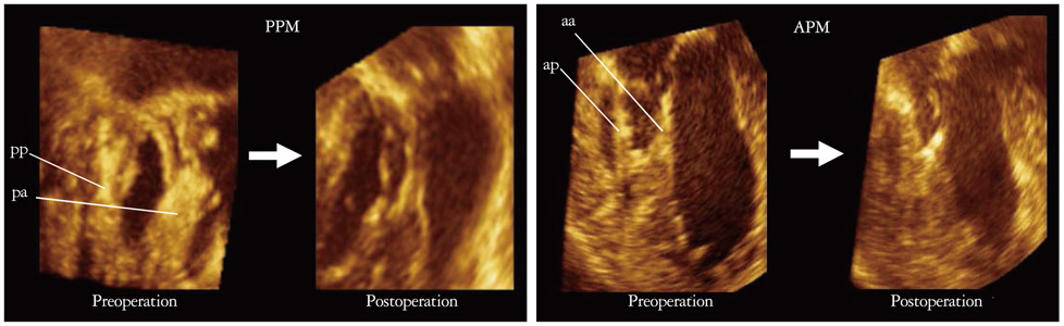
Fig. 7 Pre- and postoperative observation of papillary heads optimization method. Postoperative connected papillary muscle head for anterior leaflet and for posterior leaflet of each papillary muscle by papillary heads optimization procedure is clearly visualized by three-dimensional transesophageal echocardiography with transgastric approach. PPM: posterior papillary muscle, APM: anterior papillary muscle, pp: PPM head for posterior leaflet, pa: PPM head for anterior leaflet, aa: APM head for anterior leaflet, ap: APM head for posterior leaflet.
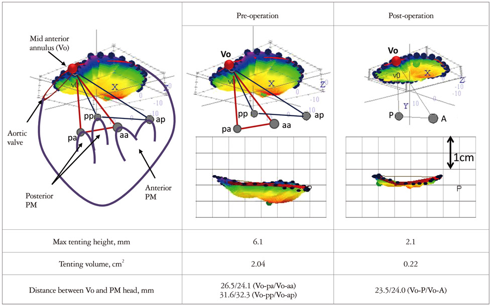
Fig. 8 Quantitative analysis of submitral structure. With the aid of quantitative software, acquired three-dimensional volume dataset allows accurate measurement of the pre and postoperative distance between papillary muscle head and mid-anterior annulus and the tenting volume, etc. Vo: mid-anterior annulus point, pp: posterior papillary muscle (PPM) head for posterior leaflet, pa: PPM head for anterior leaflet, aa: anterior papillary muscle (APM) head for anterior leaflet, ap: APM head for posterior leaflet, PM: papillary muscle.
Reference
-
1. Otto CM. Textbook of clinical echocardiography. 2009. 4th ed. Philadelphia: Saunders/Elsevier.2. Levine RA, Weyman AE, Handschumacher MD. Three-dimensional echocardiography: techniques and applications. Am J Cardiol. 1992. 69:121H–130H. discussion 131H-4H.
Article3. He S, Fontaine AA, Schwammenthal E, Yoganathan AP, Levine RA. Integrated mechanism for functional mitral regurgitation: leaflet restriction versus coapting force: in vitro studies. Circulation. 1997. 96:1826–1834.
Article4. Yiu SF, Enriquez-Sarano M, Tribouilloy C, Seward JB, Tajik AJ. Determinants of the degree of functional mitral regurgitation in patients with systolic left ventricular dysfunction: a quantitative clinical study. Circulation. 2000. 102:1400–1406.
Article5. Bursi F, Enriquez-Sarano M, Nkomo VT, Jacobsen SJ, Weston SA, Meverden RA, Roger VL. Heart failure and death after myocardial infarction in the community: the emerging role of mitral regurgitation. Circulation. 2005. 111:295–301.
Article6. Grigioni F, Enriquez-Sarano M, Zehr KJ, Bailey KR, Tajik AJ. Ischemic mitral regurgitation: long-term outcome and prognostic implications with quantitative Doppler assessment. Circulation. 2001. 103:1759–1764.
Article7. Bursi F, Barbieri A, Grigioni F, Reggianini L, Zanasi V, Leuzzi C, Ricci C, Piovaccari G, Branzi A, Modena MG. Prognostic implications of functional mitral regurgitation according to the severity of the underlying chronic heart failure: a long-term outcome study. Eur J Heart Fail. 2010. 12:382–388.
Article8. Kumanohoso T, Otsuji Y, Yoshifuku S, Matsukida K, Koriyama C, Kisanuki A, Minagoe S, Levine RA, Tei C. Mechanism of higher incidence of ischemic mitral regurgitation in patients with inferior myocardial infarction: quantitative analysis of left ventricular and mitral valve geometry in 103 patients with prior myocardial infarction. J Thorac Cardiovasc Surg. 2003. 125:135–143.
Article9. Veronesi F, Corsi C, Sugeng L, Caiani EG, Weinert L, Mor-Avi V, Cerutti S, Lamberti C, Lang RM. Quantification of mitral apparatus dynamics in functional and ischemic mitral regurgitation using real-time 3-dimensional echocardiography. J Am Soc Echocardiogr. 2008. 21:347–354.
Article10. Tsukiji M, Watanabe N, Yamaura Y, Okahashi N, Obase K, Neishi Y, Toyota E, Kawamoto T, Okura H, Ogasawara Y, Yoshida K. Three-dimensional quantitation of mitral valve coaptation by a novel software system with transthoracic real-time three-dimensional echocardiography. J Am Soc Echocardiogr. 2008. 21:43–46.
Article11. Song JM, Fukuda S, Kihara T, Shin MS, Garcia MJ, Thomas JD, Shiota T. Value of mitral valve tenting volume determined by real-time three-dimensional echocardiography in patients with functional mitral regurgitation. Am J Cardiol. 2006. 98:1088–1093.
Article12. Watanabe N, Ogasawara Y, Yamaura Y, Yamamoto K, Wada N, Kawamoto T, Toyota E, Akasaka T, Yoshida K. Geometric differences of the mitral valve tenting between anterior and inferior myocardial infarction with significant ischemic mitral regurgitation: quantitation by novel software system with transthoracic real-time three-dimensional echocardiography. J Am Soc Echocardiogr. 2006. 19:71–75.
Article13. Daimon M, Saracino G, Gillinov AM, Koyama Y, Fukuda S, Kwan J, Song JM, Kongsaerepong V, Agler DA, Thomas JD, Shiota T. Local dysfunction and asymmetrical deformation of mitral annular geometry in ischemic mitral regurgitation: a novel computerized 3D echocardiographic analysis. Echocardiography. 2008. 25:414–423.
Article14. Kim K, Kaji S, An Y, Yoshitani H, Takeuchi M, Levine RA, Otsuji Y, Furukawa Y. Mechanism of asymmetric leaflet tethering in ischemic mitral regurgitation: 3D analysis with multislice CT. JACC Cardiovasc Imaging. 2012. 5:230–232.
Article15. Saito K, Okura H, Watanabe N, Obase K, Tamada T, Koyama T, Hayashida A, Neishi Y, Kawamoto T, Yoshida K. Influence of chronic tethering of the mitral valve on mitral leaflet size and coaptation in functional mitral regurgitation. JACC Cardiovasc Imaging. 2012. 5:337–345.
Article16. Ormiston JA, Shah PM, Tei C, Wong M. Size and motion of the mitral valve annulus in man. I. A two-dimensional echocardiographic method and findings in normal subjects. Circulation. 1981. 64:113–120.
Article17. Watanabe N, Ogasawara Y, Yamaura Y, Kawamoto T, Akasaka T, Yoshida K. Geometric deformity of the mitral annulus in patients with ischemic mitral regurgitation: a real-time three-dimensional echocardiographic study. J Heart Valve Dis. 2005. 14:447–452.18. Levine RA, Handschumacher MD, Sanfilippo AJ, Hagege AA, Harrigan P, Marshall JE, Weyman AE. Three-dimensional echocardiographic reconstruction of the mitral valve, with implications for the diagnosis of mitral valve prolapse. Circulation. 1989. 80:589–598.
Article19. Kaplan SR, Bashein G, Sheehan FH, Legget ME, Munt B, Li XN, Sivarajan M, Bolson EL, Zeppa M, Arch MZ, Martin RW. Three-dimensional echocardiographic assessment of annular shape changes in the normal and regurgitant mitral valve. Am Heart J. 2000. 139:378–387.
Article20. Salgo IS, Gorman JH 3rd, Gorman RC, Jackson BM, Bowen FW, Plappert T, St John Sutton MG, Edmunds LH Jr. Effect of annular shape on leaflet curvature in reducing mitral leaflet stress. Circulation. 2002. 106:711–717.
Article21. Kwan J, Qin JX, Popović ZB, Agler DA, Thomas JD, Shiota T. Geometric changes of mitral annulus assessed by real-time 3-dimensional echocardiography: becoming enlarged and less nonplanar in the anteroposterior direction during systole in proportion to global left ventricular systolic function. J Am Soc Echocardiogr. 2004. 17:1179–1184.
Article22. Watanabe N, Ogasawara Y, Yamaura Y, Kawamoto T, Toyota E, Akasaka T, Yoshida K. Quantitation of mitral valve tenting in ischemic mitral regurgitation by transthoracic real-time three-dimensional echocardiography. J Am Coll Cardiol. 2005. 45:763–769.
Article23. Watanabe N, Ogasawara Y, Yamaura Y, Wada N, Kawamoto T, Toyota E, Akasaka T, Yoshida K. Mitral annulus flattens in ischemic mitral regurgitation: geometric differences between inferior and anterior myocardial infarction: a real-time 3-dimensional echocardiographic study. Circulation. 2005. 112:9 Suppl. I458–I462.24. Vahanian A, Baumgartner H, Bax J, Butchart E, Dion R, Filippatos G, Flachskampf F, Hall R, Iung B, Kasprzak J, Nataf P, Tornos P, Torracca L, Wenink A;. Guidelines on the management of valvular heart disease: The Task Force on the Management of Valvular Heart Disease of the European Society of Cardiology. Eur Heart J. 2007. 28:230–268.
Article25. Rankin JS, Feneley MP, Hickey MS, Muhlbaier LH, Wechsler AS, Floyd RD, Reves JG, Skelton TN, Califf RM, Lowe JE, et al. A clinical comparison of mitral valve repair versus valve replacement in ischemic mitral regurgitation. J Thorac Cardiovasc Surg. 1988. 95:165–177.
Article26. Cohn LH, Rizzo RJ, Adams DH, Couper GS, Sullivan TE, Collins JJ Jr, Aranki SF. The effect of pathophysiology on the surgical treatment of ischemic mitral regurgitation: operative and late risks of repair versus replacement. Eur J Cardiothorac Surg. 1995. 9:568–574.
Article27. Al-Radi OO, Austin PC, Tu JV, David TE, Yau TM. Mitral repair versus replacement for ischemic mitral regurgitation. Ann Thorac Surg. 2005. 79:1260–1267.
Article28. Chan V, Ruel M, Mesana TG. Mitral valve replacement is a viable alternative to mitral valve repair for ischemic mitral regurgitation: a case-matched study. Ann Thorac Surg. 2011. 92:1358–1365.
Article29. Perrault LP, Moskowitz AJ, Kron IL, Acker MA, Miller MA, Horvath KA, Thourani VH, Argenziano M, D'Alessandro DA, Blackstone EH, Moy CS, Mathew JP, Hung J, Gardner TJ, Parides MK. Optimal surgical management of severe ischemic mitral regurgitation: to repair or to replace? J Thorac Cardiovasc Surg. 2012. 143:1396–1403.
Article30. Bolling SF, Pagani FD, Deeb GM, Bach DS. Intermediate-term outcome of mitral reconstruction in cardiomyopathy. J Thorac Cardiovasc Surg. 1998. 115:381–386.
Article31. Bolling SF, Deeb GM, Bach DS. Mitral valve reconstruction in elderly, ischemic patients. Chest. 1996. 109:35–40.
Article32. Calafiore AM, Gallina S, Di Mauro M, Gaeta F, Iacò AL, D'Alessandro S, Mazzei V, Di Giammarco G. Mitral valve procedure in dilated cardiomyopathy: repair or replacement? Ann Thorac Surg. 2001. 71:1146–1152.
Article33. Zhu F, Otsuji Y, Yotsumoto G, Yuasa T, Ueno T, Yu B, Koriyama C, Hamasaki S, Biro S, Kisanuki A, Minagoe S, Levine RA, Sakata R, Tei C. Mechanism of persistent ischemic mitral regurgitation after annuloplasty: importance of augmented posterior mitral leaflet tethering. Circulation. 2005. 112:9 Suppl. I396–I401.34. Gelsomino S, Lorusso R, Caciolli S, Capecchi I, Rostagno C, Chioccioli M, De Cicco G, Billè G, Stefàno P, Gensini GF. Insights on left ventricular and valvular mechanisms of recurrent ischemic mitral regurgitation after restrictive annuloplasty and coronary artery bypass grafting. J Thorac Cardiovasc Surg. 2008. 136:507–518.
Article35. Troubil M, Marcian P, Gwozdziewicz M, Santavy P, Langova K, Nemec P, Lonsky V. Predictors of failure following restrictive annuloplasty for chronic ischemic mitral regurgitation. J Card Surg. 2012. 27:6–12.
Article36. Gelsomino S, van Garsse L, Lucà F, Lorusso R, Cheriex E, Rao CM, Caciolli S, Vizzardi E, Crudeli E, Stefàno P, Gensini GF, Maessen J. Impact of preoperative anterior leaflet tethering on the recurrence of ischemic mitral regurgitation and the lack of left ventricular reverse remodeling after restrictive annuloplasty. J Am Soc Echocardiogr. 2011. 24:1365–1375.
Article37. Magne J, Sénéchal M, Mathieu P, Dumesnil JG, Dagenais F, Pibarot P. Restrictive annuloplasty for ischemic mitral regurgitation may induce functional mitral stenosis. J Am Coll Cardiol. 2008. 51:1692–1701.
Article38. Kubota K, Otsuji Y, Ueno T, Koriyama C, Levine RA, Sakata R, Tei C. Functional mitral stenosis after surgical annuloplasty for ischemic mitral regurgitation: importance of subvalvular tethering in the mechanism and dynamic deterioration during exertion. J Thorac Cardiovasc Surg. 2010. 140:617–623.
Article39. Kainuma S, Taniguchi K, Daimon T, Sakaguchi T, Funatsu T, Kondoh H, Miyagawa S, Takeda K, Shudo Y, Masai T, Fujita S, Nishino M, Sawa Y. Osaka Cardiovascular Surgery Research (OSCAR) Group. Does stringent restrictive annuloplasty for functional mitral regurgitation cause functional mitral stenosis and pulmonary hypertension? Circulation. 2011. 124:11 Suppl. S97–S106.
Article40. Kron IL, Green GR, Cope JT. Surgical relocation of the posterior papillary muscle in chronic ischemic mitral regurgitation. Ann Thorac Surg. 2002. 74:600–601.
Article41. Hvass U, Tapia M, Baron F, Pouzet B, Shafy A. Papillary muscle sling: a new functional approach to mitral repair in patients with ischemic left ventricular dysfunction and functional mitral regurgitation. Ann Thorac Surg. 2003. 75:809–811.
Article42. Hvass U, Joudinaud T. The papillary muscle sling for ischemic mitral regurgitation. J Thorac Cardiovasc Surg. 2010. 139:418–423.
Article43. Matsui Y, Suto Y, Shimura S, Fukada Y, Naito Y, Yasuda K, Sasaki S. Impact of papillary muscles approximation on the adequacy of mitral coaptation in functional mitral regurgitation due to dilated cardiomyopathy. Ann Thorac Cardiovasc Surg. 2005. 11:164–171.44. Masuyama S, Marui A, Shimamoto T, Komeda M. Chordal translocation: secondary chordal cutting in conjunction with artificial chordae for preserving valvular-ventricular interaction in the treatment of functional mitral regurgitation. J Heart Valve Dis. 2009. 18:142–146.45. Fattouch K, Murana G, Castrovinci S, Mossuto C, Sampognaro R, Borruso MG, Bertolino EC, Caccamo G, Ruvolo G, Lancellotti P. Mitral valve annuloplasty and papillary muscle relocation oriented by 3-dimensional transesophageal echocardiography for severe functional mitral regurgitation. J Thorac Cardiovasc Surg. 2012. 143:4 Suppl. S38–S42.
Article46. Obase K, Komeda M, Okura H, Yoshida K. A new echocardiographic window to visualize the mitral valve complex during mitral valve repair for functional mitral regurgitation. J Thorac Cardiovasc Surg. 2012. 143:e42–e44.
Article
- Full Text Links
-
- Actions
-
Cited
- CITED
-
- Close
- Share
-
- Similar articles
-
- Echocardiographic Assessment of Mitral Valve Regurgitation
- A Modified Echocardiographic Classification of Mitral Valve Regurgitation Mechanism: The Role of Three-dimensional Echocardiography
- Assessment of Mitral Stenosis by Doppler Echocardiography: Influence of Regurgitation on Doppler Pressure Half-Time
- Congenital Double-Orifice Mitral Valve with Mitral Valve Prolapse and Severe Mitral Regurgitation
- Mitral Valve Repair
