Two Cases of Incidentally Diagnosed Idiopathic Left Atrial Appendage Ostial Stenosis
- Affiliations
-
- 1Division of Cardiology, Korea University Cardiovascular Center, Korea University Anam Hospital, Seoul, Korea. wjshimmd@unitel.co.kr
- KMID: 2177311
- DOI: http://doi.org/10.4250/jcu.2010.18.3.112
Abstract
- We report here on 2 cases of idiopathic left atrial appendage ostial stenosis (LAA), and this rare finding was detected on transesophageal echocardiography. Its clinical implication is still unknown, given the small number of reported cases. Incompletely ligated LAA has characteristics similar to those observed in idiopathic LAA ostial stenosis, including the narrowed orifice, the small LAA cavity and the accelerated blood flow across the stenotic area. Since the incompletely ligated LAA has been reported to be complicated with thromboembolic events, we can assumed that the patients with idiopathic LAA ostial stenosis have a higher risk of thromboembolism than those with a normal LAA structure.
Keyword
MeSH Terms
Figure
-
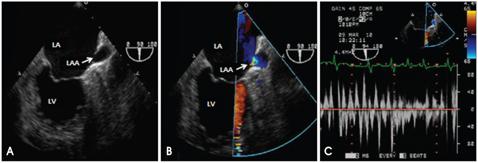
Fig. 1 Transesophageal echocardiography of the case 1 revealed a small tubular shaped left atrial appendage with a narrowed orifice, and the maximal diameter of the orifice was only 4.8 mm (A). Doppler examination showed significant flow acceleration across the stenotic area with a peak velocity more than 100 cm/sec (B and C). LA: left atrium, LAA: left atrial appendage, LV: left ventricle.
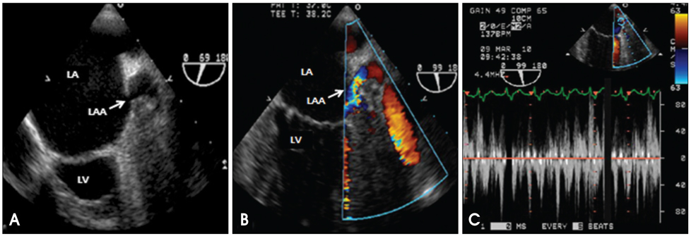
Fig. 2 Transesophageal echocardiography of the case 2 revealed a long tubular shaped left atrial appendage with a narrowed orifice, and the maximal diameter of the orifice was only 3.8 mm (A). The peak velocity across the narrowed orifice measured more than 110 cm/sec with flow acceleration on Doppler echocardiography (B and C). LA: left atrium, LAA: left atrial appendage, LV: left ventricle.
Reference
-
1. Veinot JP, Harrity PJ, Gentile F, Khandheria BK, Bailey KR, Eickholt JT, Seward JB, Tajik AJ, Edwards WD. Anatomy of the normal left atrial appendage: a quantitative study of age related changes in 500 autopsy hearts: implications for echocardiographic examination. Circulation. 1997. 96:3112–3115.2. Coughlan B, Lang RM, Spencer KT. Left atrial appendage stenosis. J Am Soc Echocardiogr. 1999. 12:882–883.
Article3. Stern JD, Skolnick AH, Freedberg RS, Kronzon I. Isolated left atrial appendage ostial stenosis. Eur J Echocardiogr. 2009. 10:702–703.
Article4. Rosenzweig BP, Katz E, Kort S, Schloss M, Kronzon I. Thromboembolus from a ligated left atrial appendage. J Am Soc Echocardiogr. 2001. 14:396–398.
Article5. Oneglia C, Muneretto C, Rusconi C. Transesophageal investigation of surgically ligated left atrial appendage. Echocardiography. 2004. 21:617–619.
Article6. Katz ES, Tsiamtsiouris T, Applebaum RM, Schwartzbard A, Tunick PA, Kronzon I. Surgical left atrial appendage ligation is frequently incomplete: a transesophageal echocardiographic study. J Am Coll Cardiol. 2000. 36:468–471.
Article7. Kanderian AS, Gillinov AM, Pettersson GB, Blackstone E, Klein AL. Success of surgical left atrial appendage closure: assessment by transesophageal echocardiography. J Am Coll Cardiol. 2008. 52:924–929.8. Cha TJ, Lee CH, Kim HJ, Lee YS, Jung HG, Choi H, Joo SJ, Lee JW. Assessment of left atrial appendage flow pattern using multiplane transesophageal echocardiography in patients with nonrheumatic atrial fibrillation and ischemic stroke. J Korean Soc Echocardiogr. 1997. 5:103–114.
Article
- Full Text Links
-
- Actions
-
Cited
- CITED
-
- Close
- Share
-
- Similar articles
-
- Coronary Neovascularity and Fistula Formation in Left Atrial Thrombosis
- Percutaneous Mitral Balloon Valveloplasty after Successful Resolution of Left Atrial Appendage Thrombi by Oral Anticoagulation
- Persistent Atrial Fibrillation Related to a Congenital Pericardial Defect and Left Atrial Appendage Herniation
- The Influence of Electrical Cardioversion for Atrial Fibrillation on Left Atrial Appendage Function: A Transesophageal Echocardiography Study
- Free-Floating Left Atrial Thrombus with Recurrent cerebral Embolic Event Associated Mitral Stenosis
