Short-term evaluation of dental implants in a diabetic population: an in vivo study
- Affiliations
-
- 1Department of Prosthodontics, Faculty of Dental Sciences, Sri Ramachandra University, Porur, Chennai, India. athiban_mds@rediffmail.com
- KMID: 2176407
- DOI: http://doi.org/10.4047/jap.2012.4.3.134
Abstract
- PURPOSE
The study was conducted to evaluate the efficacy of implant supported tooth replacement in diabetic patients.
MATERIALS AND METHODS
The study involved placement of implants (UNITI implants, Equinox Medical Technologies, Zeist, Holland, diameter of 3.7 mm and length 13 mm) in five diabetic patients (three females and two males) of age ranging from 35-65 years with acceptable metabolic control of plasma glucose. All patients included in the study were indicated for single tooth maxillary central incisor replacement, with the adjacent teeth intact. The survival of the restored implants was assessed for a period of three months by measurement of crestal bone heights, bleeding on probing and micro flora predominance. Paired t-test was done to find out the difference in the microbial colonization, bleeding on probing and crestal bone loss. P values of less than 0.05 were taken to indicate statistical significance.
RESULTS
Results indicated that there was a significant reduction in bleeding on probing and colonization at the end of three months and the bone loss was not statistically significant.
CONCLUSION
The study explores the hypothesis that patients with diabetes are appropriate candidates for implants and justifies the continued evaluation of the impact of diabetes on implant success and complications.
Keyword
MeSH Terms
Figure
-
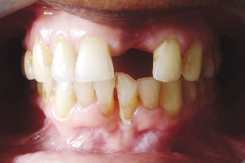
Fig. 1 Preoperative facial view of single missing maxillary central incisor.
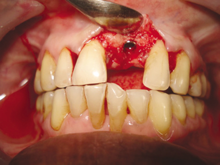
Fig. 2 UNITI implant (Equinox Medical Technologies, Zeist, Holland) with a diameter of 3.7 mm and length 13 mm placed.
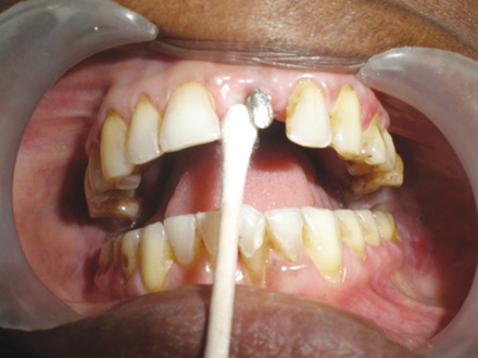
Fig. 3 Specimen collection using sterile swab to identify microorganisms located subgingivally along the implant site.

Fig. 4 Transfer of samples to vials.
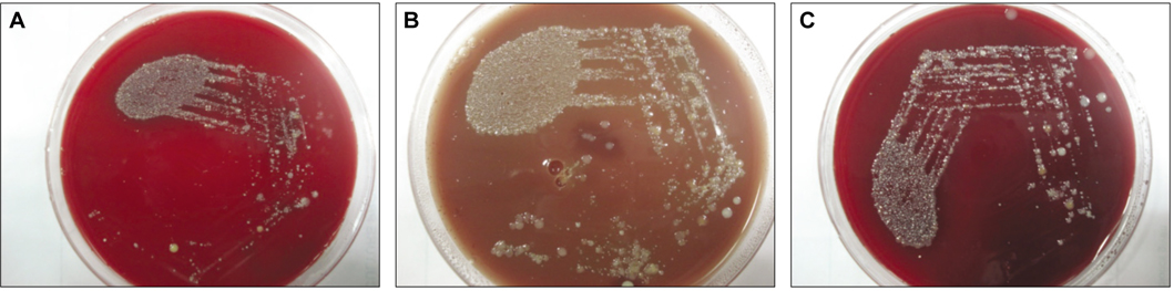
Fig. 5 A: Blood agar medium demonstrating colonization of fastidious organisms, B: Chocolate agar medium showing largest proportion of microorganisms obtained from the samples. This is a non-selective medium that showed colonies of respiratory bacteria, anaerobic organisms and gram positive cocci, C: MacConkey agar medium showing colonization. Colonies of Gram negative bacteria were seen.
Reference
-
1. Kotsovilis S, Karoussis IK, Fourmousis I. A comprehensive and critical review of dental implant placement in diabetic animals and patients. Clin Oral Implants Res. 2006. 17:587–599.2. Balshi TJ, Wolfinger GJ. Dental implants in the diabetic patient: a retrospective study. Implant Dent. 1999. 8:355–359.3. Azza Ezz El-Arab , Zahran A. Osseo integrated implants for single tooth replacement in type 1 diabetic patients: a 2 year follow up study. Cairo Dental J. 2000. 16:339–346.4. Leonhardt A, Dahlén G, Renvert S. Five-year clinical, microbiological, and radiological outcome following treatment of peri-implantitis in man. J Periodontol. 2003. 74:1415–1422.5. Peled M, Ardekian L, Tagger-Green N, Gutmacher Z, Machtei EE. Dental implants in patients with type 2 diabetes mellitus: a clinical study. Implant Dent. 2003. 12:116–122.6. Al Jabbari Y, Nagy WW, Iacopino AM. Implant dentistry for geriatric patients: a review of the literature. Quintessence Int. 2003. 34:281–285.7. Mombelli A, van Oosten MA, Schurch E Jr, Land NP. The microbiota associated with successful or failing osseointegrated titanium implants. Oral Microbiol Immunol. 1987. 2:145–151.8. Cukovic-Bagic I, Verzak Z, Car N, Car A. Tooth loss among diabetic patients. Diabetologia Croatica. 2004. 33:23–27.9. Casap N, Nimri S, Ziv E, Sela J, Samuni Y. Type 2 diabetes has minimal effect on osseointegration of titanium implants in Psammomys obesus. Clin Oral Implants Res. 2008. 19:458–464.10. Balshi SF, Wolfinger GJ, Balshi TJ. An examination of immediately loaded dental implant stability in the diabetic patient using resonance frequency analysis (RFA). Quintessence Int. 2007. 38:271–279.11. Leonhardt A, Dahlén G, Renvert S. Five-year clinical, microbiological, and radiological outcome following treatment of peri-implantitis in man. J Periodontol. 2003. 74:1415–1422.12. Salvi GE, Fürst MM, Lang NP, Persson GR. One-year bacterial colonization patterns of Staphylococcus aureus and other bacteria at implants and adjacent teeth. Clin Oral Implants Res. 2008. 19:242–248.
- Full Text Links
-
- Actions
-
Cited
- CITED
-
- Close
- Share
-
- Similar articles
-
- Recent advances in dental implants
- Treatment concepts for the posterior maxilla and mandible: short implants versus long implants in augmented bone
- Outcomes of dental implant treatment in patients with generalized aggressive periodontitis: a systematic review
- A short-term clinical study of marginal bone level change around microthreaded and platform-switched implants
- Interrelationship between periodontal parameters for the evaluation of clinically stable dental implants
