Effect of calcium hydroxide on bond strength of dentin bonding systems
- Affiliations
-
- 1Department of Conservative Dentistry, Division of Dentistry, Graduate of Kyung Hee University, Korea. psangjin@khu.ac.kr
- KMID: 2175870
- DOI: http://doi.org/10.5395/JKACD.2007.32.3.198
Abstract
- The purpose of this study was to investigate the effect of calcium hydroxide on dentin bonding strength of various dentin bonding systems as a function of time in composite resin restoration. Dentin adhesives used in this study were Scotchbond Multipurpose, Single Bond, SE Bond and Prompt L-Pop. Flat dentin surfaces adjacent to pulp chamber were created, then Ca(OH)2 and saline were mixed and applied on dentin surface of experimental group, then IRM was used to cover the mixture on dentin surface and the specimens were stored at 36.5degrees C for experiment period (7 days, 30 days). After removing IRM and Ca(OH)2, each dentin adhesives were treated on dentin surfaces. Composite resin (Z-250, 3M) was placed with 5 mm height and was light-cured for 20 seconds. After stored in distilled water for 24 hours, each dentin-composite bonded spicemen was embedded in epoxy resin and sectioned into 1.0 x 1.0 mm2 cross section composite-dentin beams. Specimen was mounted on zig of Universal testing machine and microTBS test was performed. SEM analysis was performed to examine the fractured surfaces. The results suggested that applying calcium hydroxide did not show significant difference in dentin bonding strength.
Keyword
MeSH Terms
Figure
-
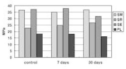
Figure 2 Micro-tensile bond strength of 12 experimental groups.
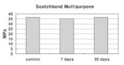
Figure 3 Micro-tensile bond strength in group of SM.

Figure 4 Micro-tensile bond strength in group of SB.
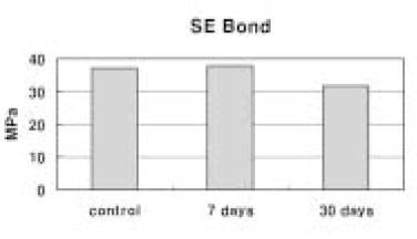
Figure 5 Micro-tensile bond strength in group of SE.
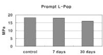
Figure 6 Micro-tensile bond strength in group of PL.

Figure 7 SEM photograph of the fractured surface of SM/C group, showing cohesive failure. The failure occurred at the bottom of the hybrid layer and there are resin tags in the dentinal tubules that fractured at the bottom of the hybrid layer.

Figure 8 SEM photograph of the fractured surface of SM/30 group, showing cohesive failure. The failure occurred at the bottom of the hybrid layer and there are resin tags in the dentinal tubules that fractured at the bottom of the hybrid layer.
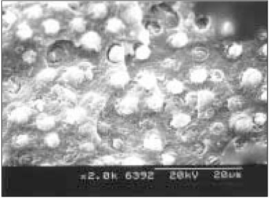
Figure 9 SEM photograph of the fractured surface of SB/C group, showing mixed failure. The failure occurred both at the top of the hybrid layer and in the bottom of the hybrid layer.
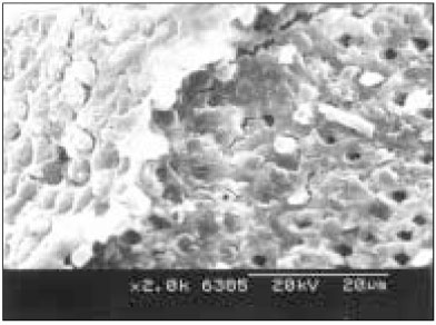
Figure 10 SEM photograph of the fractured surface of SB/30 group, showing mixed failure. The failure occurred both at the top of the hybrid layer and in the bottom of the hybrid layer.
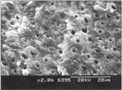
Figure 11 SEM photograph of the fractured surface of SE/C group, showing cohesive failure. The failure occurred at the bottom of the hybrid layer and exposed dentin, which was not enveloped by resin.

Figure 12 SEM photograph of the fractured surface of SE/30 group, showing cohesive failure. The failure occurred at the bottom of the hybrid layer and exposed dentin, which was not enveloped by resin.

Figure 13 SEM photograph of the fractured surface of PL/C group, showing adhesive failure. The failure occurred between the resin and the top of the hybrid layer.
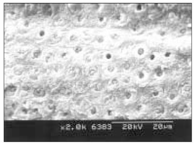
Figure 14 SEM photograph of the fractured surface of PL/30 group, showing adhesive failure. The failure occurred between the resin and the top of the hybrid layer.
Reference
-
1. Orstavik D. Antibacterial properties of endodontic materials. Int Endod J. 1988. 21:161–169.
Article2. Chong BS, Pitt Ford TR. The role of intracanal medication in root canal treatment. Int Endod J. 1992. 25(2):97–106.
Article3. Unemori M, et al. Composite resin restoration and postoperative sensitivity: clinical follow-up in an undergraduate program. J Dent. 2001. 29(1):7–13.
Article4. Ulusu T, et al. Comparison of the effect of insertion techniques of a resin composite on dentinal adaptation of two visible light-cured bases: direct evaluation versus a replica technique. Quintessence Int. 1996. 27(1):63–68.5. Cox CF, Suzuki S. Re-evaluating pulp protection: calcium hydroxide liners vs. cohesive hybridization. J Am Dent Assoc. 1994. 125(7):823–831.
Article6. Suliman AA, Chan KC. Microleakage between different types of base materials. J Prosthet Dent. 1992. 67(2):153–156.
Article7. Staehle HJ, et al. The marginal sealing of composite inlays with different cavity liners. Schweiz Monatsschr Zahnmed. 1992. 102(10):1189–1194.8. Fitzgerald M, Heys RJ. A clinical and histological evaluation of conservative pulpal therapy in human teeth. Oper Dent. 1991. 16(3):101–112.9. Papadakou M, et al. Adaptation of two different calcium hydroxide bases under a composite restoration. J Dent. 1990. 18(5):276–280.
Article10. Peliz MI, et al. Scanning electron microscope analysis of internal adaptation of materials used for pulp protection under composite resin restorations. J Esthet Restor Dent. 2005. 17(2):118–128.
Article11. Ersin NK, Eronat N. The comparison of a dentin adhesive with calcium hydroxide as a pulp-capping agent on the exposed pulps of human and sheep teeth. Quintessence Int. 2005. 36(4):271–280.12. Subay RK, Demirci M. Pulp tissue reactions to a dentin bonding agent as a direct capping agent. J Endod. 2005. 31(3):201–204.
Article13. Scarano A, et al. A Direct capping with four different materials in humans: histological analysis of odontoblast activity. J Endod. 2003. 29(11):729–734.
Article14. Cox CF, et al. Biocompatibility of surface-sealed dental materials against exposed pulps. J Prosthet Dent. 1987. 57(1):1–8.
Article15. Foreman PC, Barnes IE. A review of calcium hydroxide. Int Endod J. 1990. 23:283–297.
Article16. Eidelman E, et al. Remineralization of carious dentin treated with calcium hydroxide. J Dent Child. 1965. 32(4):218–225.17. Kehoe JC. pH reversal following in vitro bleaching of pulpless teeth. J Endod. 1987. 13(1):6–9.18. de Oliveira LD, et al. Sealing evaluation of the cervical base in intracoronal bleaching. Dent Traumatol. 2003. 19(6):309–313.
Article19. Lambrianidis T, et al. Effect of calcium hydroxide as a supplementary barrier in the radicular penetration of hydrogen peroxide during intracoronal bleaching in vitro. Int Endod J. 2002. 35(12):985–990.
Article20. Andreasen JO, Kristerson L. The effect of extra-alveolar root filling with calcium hydroxide on periodontal healing after replantation of permanent incisors in monkeys. J Endod. 1981. 7(8):349–354.
Article21. Chung HA, et al. Adhesion of glass-ionomer cement sealers to bovine dentin conditioned with intracanal medications. J Endod. 2001. 27(2):85–88.
Article22. Windley W III, et al. The effect short-term calciun hydroxide treatment on dentin bond strength to composite resin. Dent Traumatol. 2003. 19(2):79–84.
Article23. Shono Y, et al. Regional measurement of resin-dentin bonding as an array. J Dent Res. 1999. 78(2):699–705.
Article24. Tay FR, Pashley DH. Aggressiveness of contemporary self-etching systems. I: Depth of penetration beyond dentin smear layers. Dent Mater. 2001. 17(4):296–308.25. Jang YI, Choi KK, Park SJ. Compatibility between two step self-etching adhesives and composites with different curing mode. J Korean Acad Conserv Dent. 2006. 31:(in process).26. Tanumiharja M, et al. Microtensile bond strengths of seven dentin adhesive systems. Dent Mater. 2000. 16:180–187.
Article27. Andreasen JO, Kristenson L. Long-term calcium hydroxide as a root canal dressing may increase risk of root fracture. Dent Traumatol. 2002. 18:134–137.
Article28. Pioch T, et al. The nanoleakage phenomenon: influence of moist vs dry bonding. J Adhes Dent. 2002. 4:23–30.29. Swift EJ Jr. Dentin bonding: what is the state of the art? Compend Contin Educ Dent. 2001. 22:12 Suppl. 4–7. quiz 18.30. Perdigao J. Dentin bonding as a function of dentin structure. Dent Clin North Am. 2002. 46:277–301.
Article
- Full Text Links
-
- Actions
-
Cited
- CITED
-
- Close
- Share
-
- Similar articles
-
- The effect of bonding resin on bond strength of dual-cure resin cements
- Comparative evaluation of micro-shear bond strength between two different luting methods of resin cement to dentin
- The effect of repeated bonding on the shear bond strength of different resin cements to enamel and dentin
- Effect of calcium hydroxide application time on dentin
- The comparison of microtensile bond strength with immediate and delayed dentin sealing
