J Korean Acad Conserv Dent.
2004 Nov;29(6):504-514. 10.5395/JKACD.2004.29.6.504.
Dentin bond strength of bonding agents cured with light emitting diode
- Affiliations
-
- 1Department of Conservative Dentistry, College of Dentistry, Seoul National University, Korea. umcmoon@snu.ac.kr
- KMID: 2175651
- DOI: http://doi.org/10.5395/JKACD.2004.29.6.504
Abstract
- This study compared the dentin shear bond strengths of currently used dentin bonding agents that were irradiated with an LED (Elipar FreeLight, 3M-ESPE) and a halogen light (VIP, BISCO). The optical characteristics of two light curing units were evaluated. Extracted human third molars were prepared to expose the occlusal dentin and the bonding procedures were performed under the irradiation with each light curing unit. The dentin bonding agents used in this study were Scotchbond Multipurpose (3M ESPE), Single Bond (3M ESPE), One-Step (Bisco), Clearfil SE bond (Kuraray), and Adper Prompt (3M ESPE). The shear test was performed by employing the design of a chisel-on-iris supported with a Teflon wall. The fractured dentin surface was observed with SEM to determine the failure mode. The spectral appearance of the LED light curing unit was different from that of the halogen light curing unit in terms of maximum peak and distribution. The LED LCU (maximum peak in 465 nm) shows a narrower spectral distribution than the halogen LCU (maximum peak in 487 nm). With the exception of the Clearfil SE bond (P < 0.05), each 4 dentin bonding agents showed no significant difference between the halogen light-cured group and the LED light-cured group in the mean shear bond strength (P > 0.05). The results can be explained by the strong correlation between the absorption spectrum of camphoroquinone and the narrow emission spectrum of LED.
MeSH Terms
Figure
-
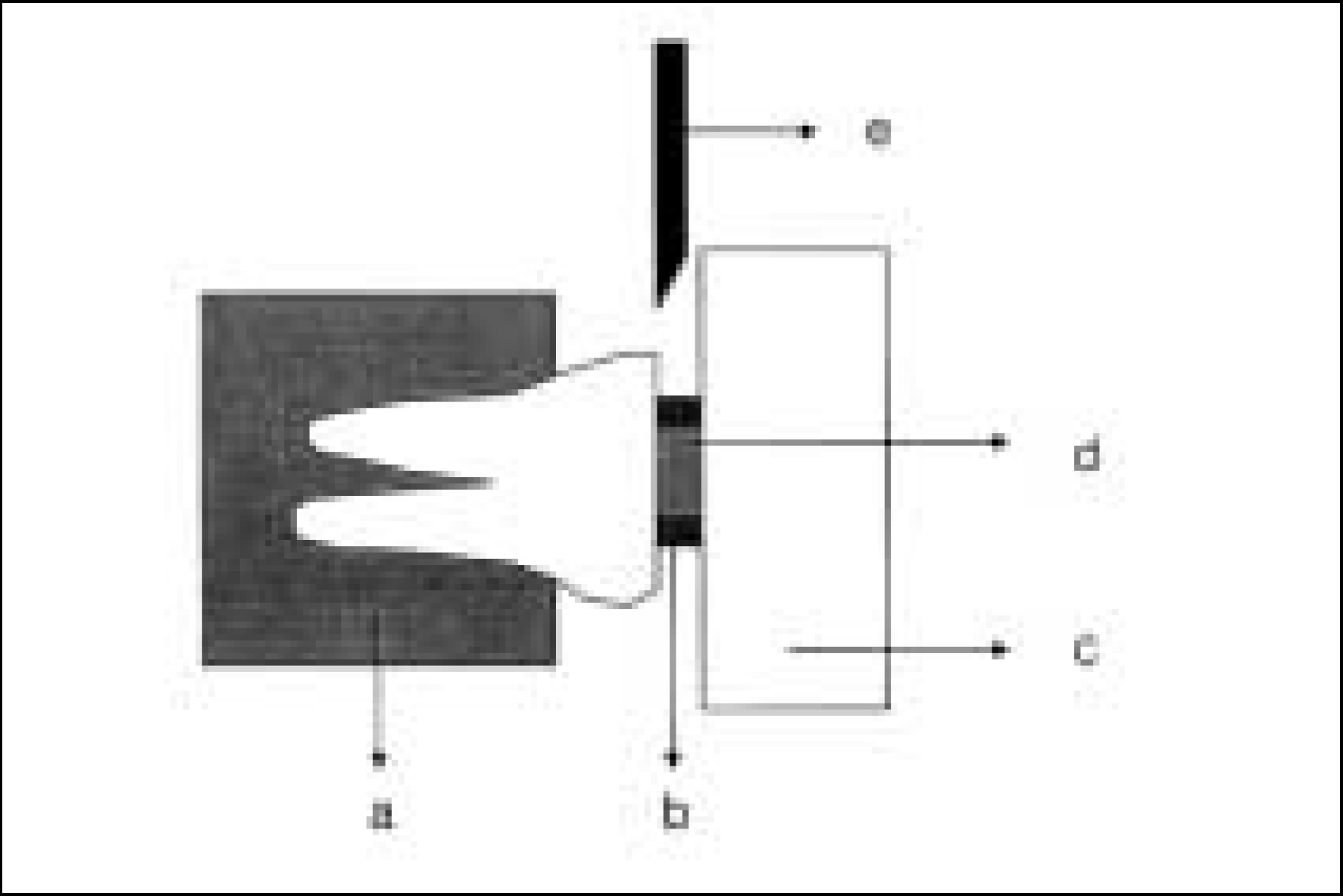
Figure 1. Schematic diagram of the shear bond strength test used in this study. Embedding resin Teflon-coated metal iris Teflon wall Composite Load application chisel
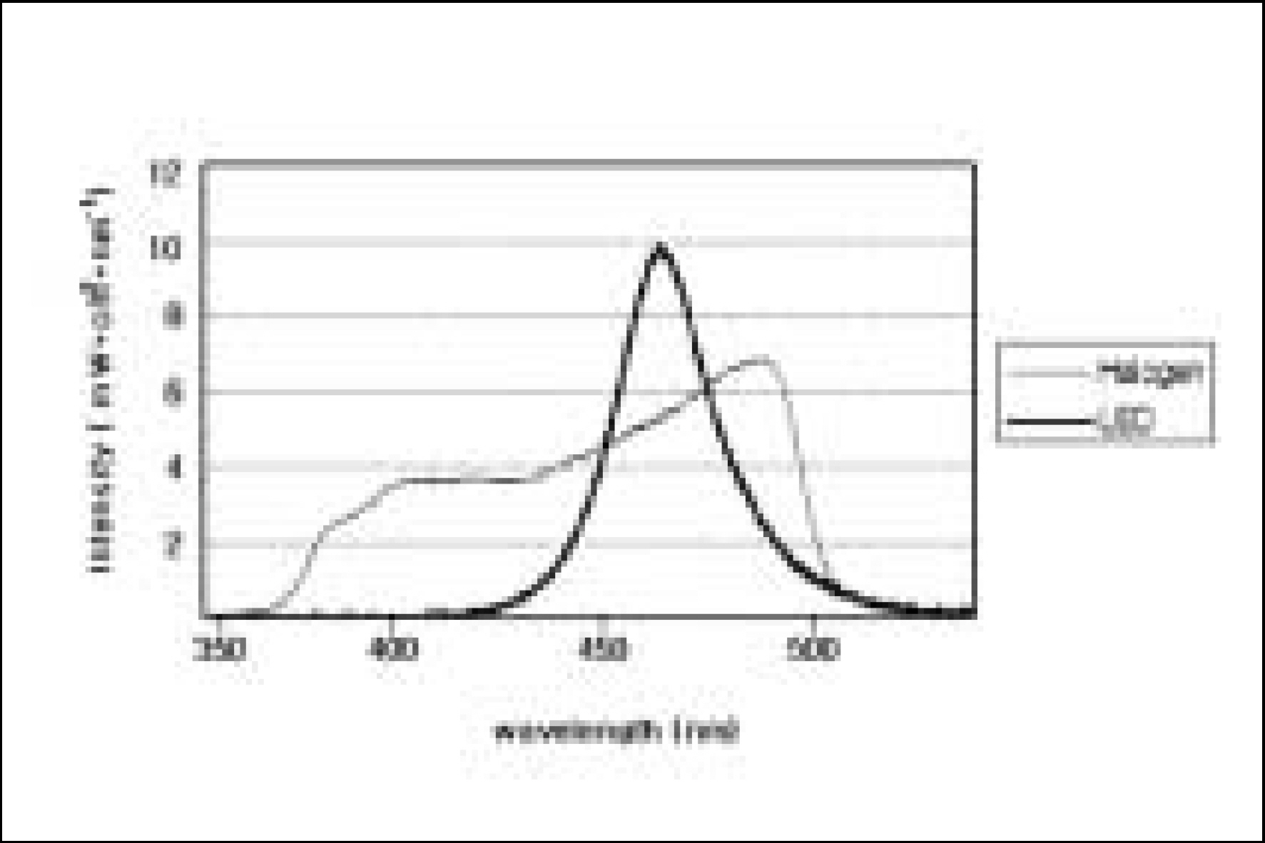
Figure 2. Spectral distributions of the two LCUs.
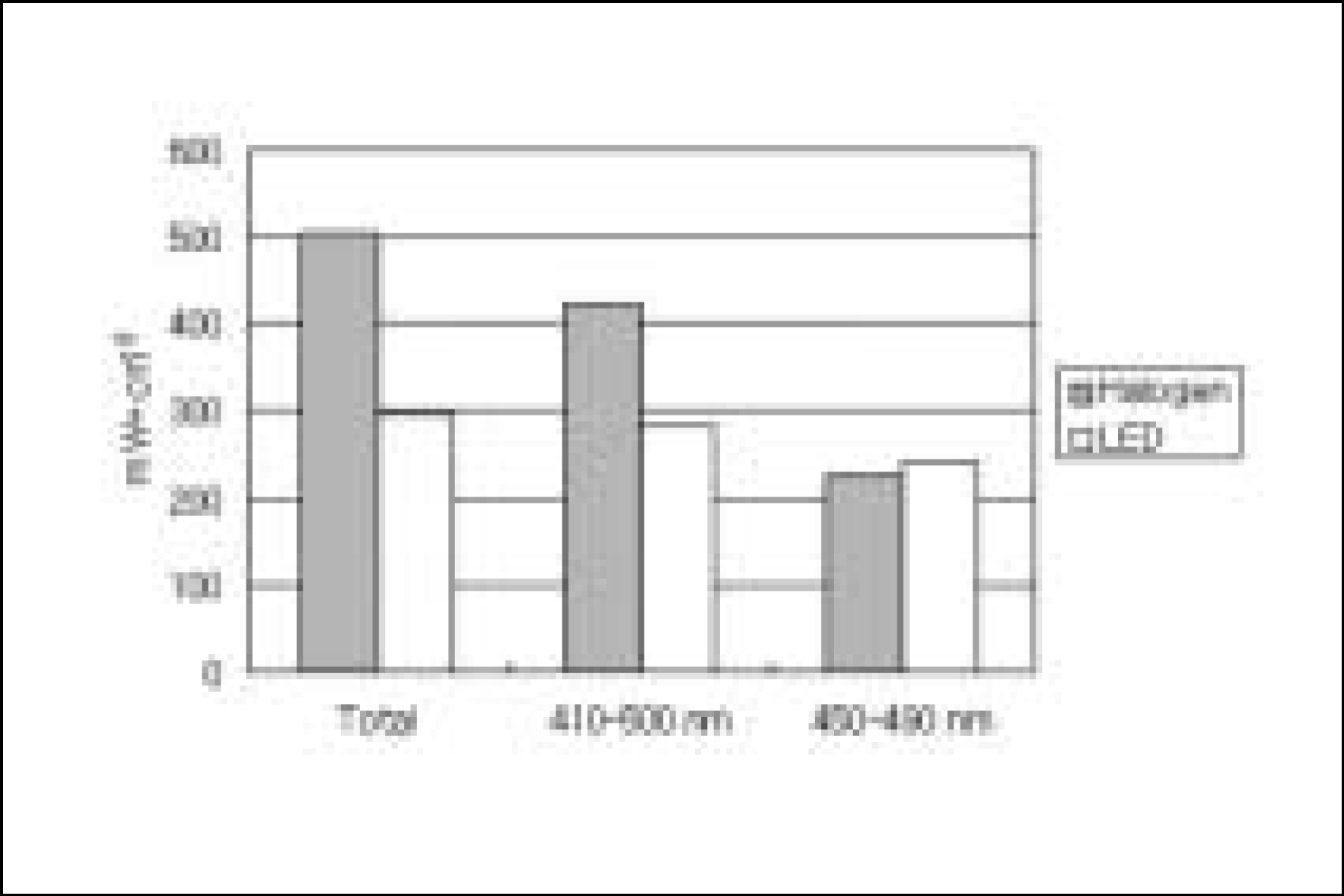
Figure 3. Spectral intensity of two LCUs used in this study.
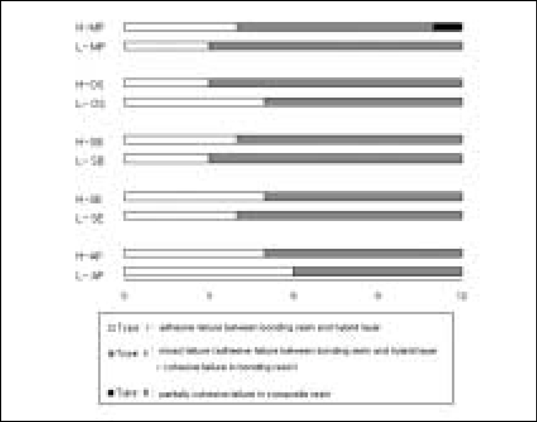
Figure 4. The failure mode.
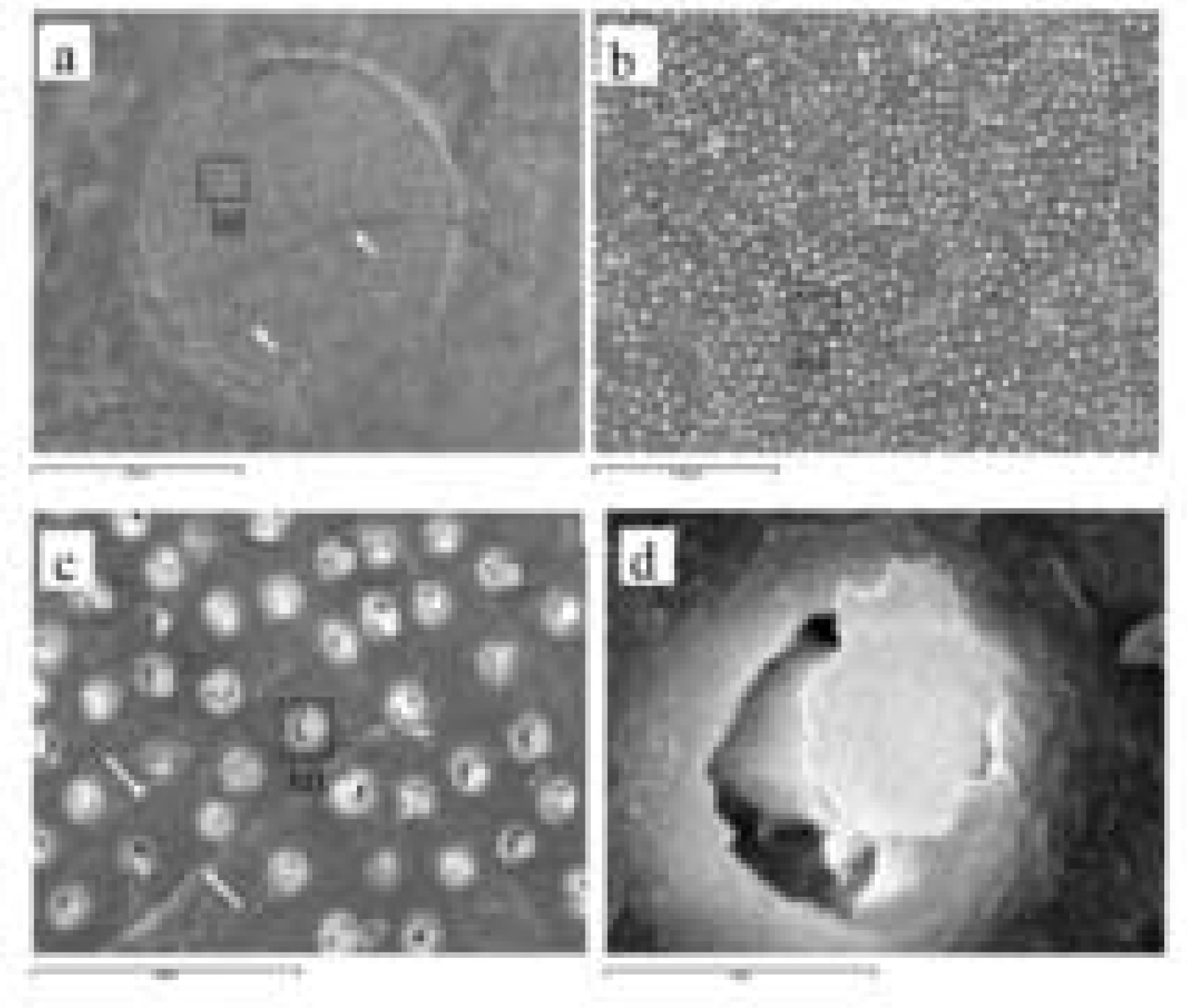
Figure 5. Adhesive failure.
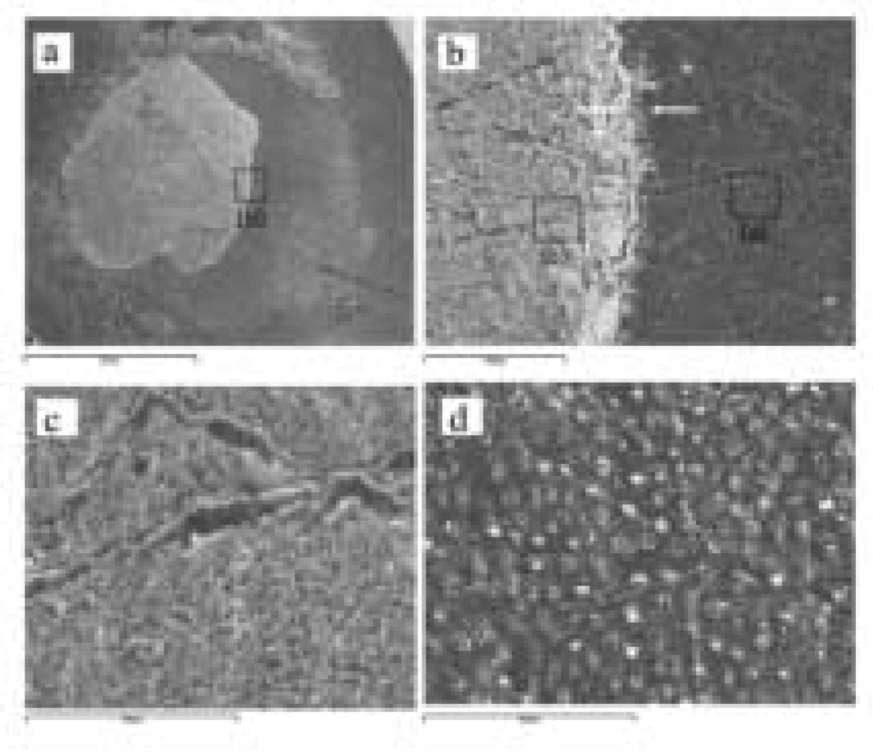
Figure 6. Mixed failure; adhesive failure + cohesive failure within the DBA.
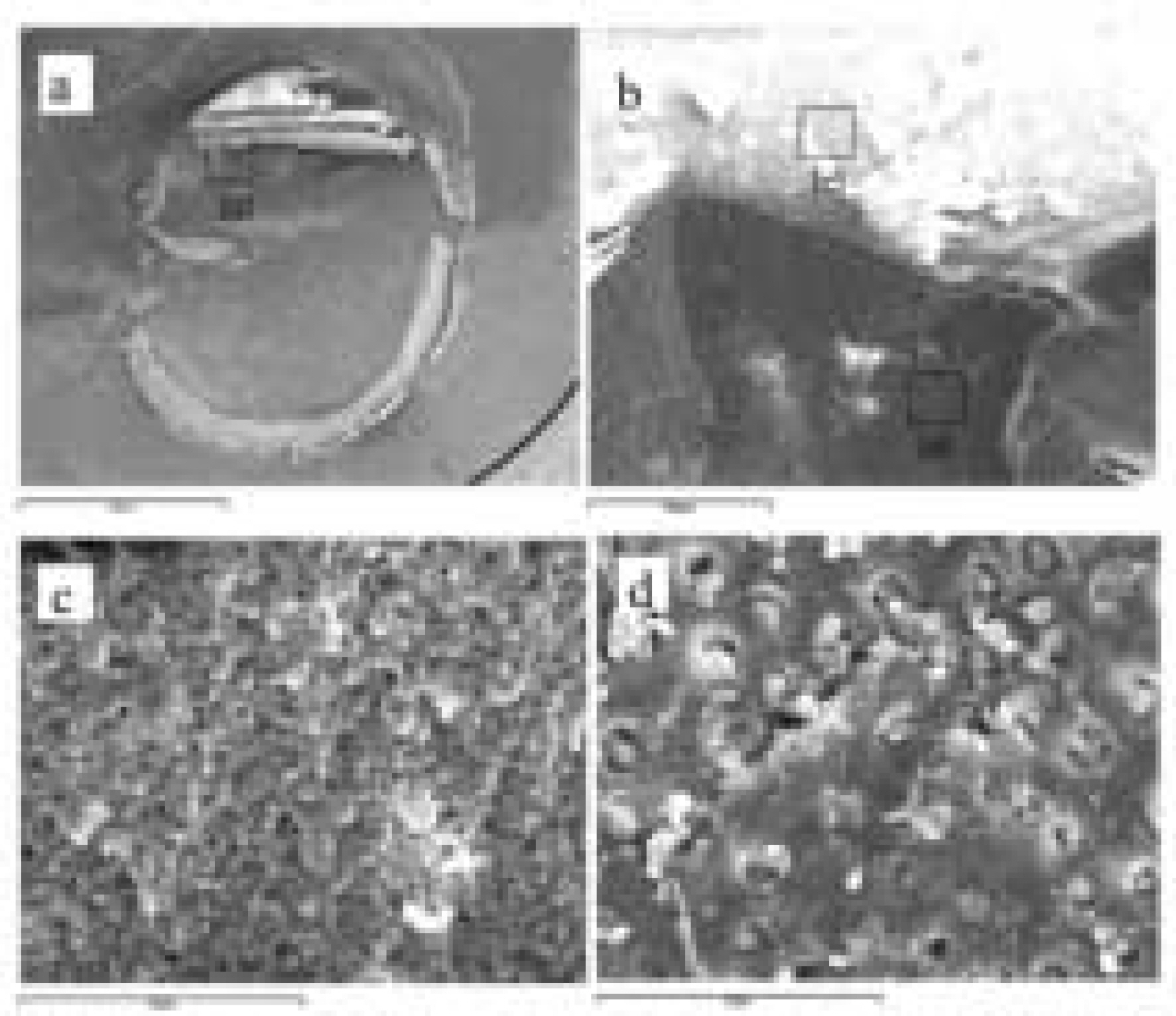
Figure 7. Partially cohesive failure within the composite.
Cited by 1 articles
-
Effect of infection control barrier thickness on light curing units
Hoon-Sang Chang, Seok-Ryun Lee, Sung-Ok Hong, Hyun-Wook Ryu, Chang-Kyu Song, Kyung-San Min
J Korean Acad Conserv Dent. 2010;35(5):368-373. doi: 10.5395/JKACD.2010.35.5.368.
Reference
-
References
1. Council on Dental Materials, Instruments, and Equipment. Visible light-cured and activating units. J Am Dent Assoc. 110(1):100–102. 1985.2. Blankenau RJ, Kelsey WP, Powell GL, Shearer GO, Barkmeier WW, Cavel WT. Degree of composite resin polymerization with visible light and argon laser. Am J Dent. 4(1):40–42. 1991.3. Ferracane JL, Mitchem JC, Condon JR, Todd R. Wear and marginal breakdown of composites with various degrees of cure. J Dent Res. 76(8):1508–1516. 1997.
Article4. Rueggeberg FA, Twiggs SW, Caughman WF, Khajotia S. Life-time intensity profiles of 11 light-curing units. J Dent Res. 75:380 Abstr.. (No. 2897):1996.5. Friedman J. Variability of lamp characteristics in dental curing lights. J Esthet Dent. 1(6):189–190. 1989.
Article6. Martin FE. A survey of the efficiency of visible light curing units. J Dent. 26(3):239–243. 1998.
Article7. Barghi N, Berry T, Hatton C. Evaluating intensity output of curing lights in private dental offices. J Am Dent Assoc. 125(7):992–996. 1994.
Article8. Miyazaki M, Hattori T, Ichiishi Y, Kondo M, Onose H, Moore BK. Evaluation of curing units used in private dental offices. Oper Dent. 23(2):50–54. 1998.9. Mills RW. Blue light emitting diodes: Another method of light curing? Br Dent J. 178:169. 1995.10. Nakamura S, Mukai T, Senoh M. Candela-class high brightness InGaN/AlGaN double heterostructure bluelight-emitting diodes. Appl Phys Lett. 64:1687–1689. 1994.11. Duke ES. Light-emitting diodes in composite resin photopolymerization. Compend Contin Educ Dent. 22(9):722–725. 2001.12. Yap AU, Soh MS. Thermal emission by different light-curing units. Oper Dent. 28(3):260–266. 2003.13. Fujibayashi K, Ishimaru K, Takahashi N, Kohno A. Newly developed curing unit using blue light-emitting diodes. Dent Jap. 34:49–53. 1998.14. Mills RW, Jandt KD, Ashworth SH. Dental composite depth of cure with halogen and blue light emitting diode (LED) technology. Br Dent J. 186(8):388–391. 1999.15. Stahl F, Ashworth SH, Jandt KD, Mills RW. Light-emitting diode (LED) polymerization of dental composites: flexural properties and polymerization potential. Biomaterials. 21(13):1379–1385. 2000.16. Mills RW, Uhl A, Jandt KD. Optical power outputs, spectral and dental composite depths of cure, obtained with blue light emitting diode (LED) and halogen light curing units (LCUs). Br Dent J. 193(8):459–463. 2002.17. Hansen EK, Asumussen E. Reliability of three dental radiometers. Scand J Dent Res. 101(2):115–119. 1993.18. Leonard DL, Charlton DG, Hilton TJ. Effect of curing-tip diameter on the accuracy of dental radiometers. Oper Dent. 24(1):31–37. 1999.19. Cook WD. Spectral distribution of dental photopolymerization sources. J Dent Res. 61:1436–1438. 1982.20. McCabe JF, Carnick TE. Output from visible-light activation units and depth of cure of light-activated composites. J Dent Res. 68(11):1534–1539. 1989.
Article21. Nomoto R. Effect of light wavelength on polymerization of light-cured resins. Dent Mater J. 16(1):60–73. 1997.
Article22. Dickens SH, Milos MF. Relationship of dentin shear bond strengths to different laboratory test designs. Am J Dent. 15(3):185–192. 2002.23. Versluis A, Tantbirojn D, Douglas WH. Why do shear bond tests pull out dentin? J Dent Res. 76(6):1298–1307. 1997.
Article24. Stansbury JW. Curing dental resins and composites by photopolymerization. J Esthet Dent. 12:300–308. 2000.
Article25. Hayakawa T, Kikutake K, Nemoto K. Effectiveness of the addition of water-soluble photoinitiator into the self-etching primers on the adhesion of a resin composite to polished dentin and enamel. Dent Mater J. 18(3):324–333. 1999.
Article26. Burgess JO, DeGoes M, Walker R, Ripps AH. An evaluation of four light-curing units comparing soft and hard curing. Pract Periodontics Aesthet Dent. 11(1):125–132. 1999.27. Koliniotou-Kubia E, Jacobsen PH. The effect of irradi-antion time on the physicalperoperties of light-cured resins. Clin Mater. 6(1):21–28. 1990.28. Takahashi A, Sato Y, Uno S, Pereira PN, Sano H. Effects of mechanical properties of adhesive resins on bond strength to dentin. Dent Mater. 18(3):263–268. 2002.
Article
- Full Text Links
-
- Actions
-
Cited
- CITED
-
- Close
- Share
-
- Similar articles
-
- The shear bond strength and adhesive failure pattern in bracket bonding with different light-curing methods
- Shear bond strength of dentin bonding agents cured with a Plasma Arc curing light
- Shear bond strength and failure patterns according to the material of resin base in indirect racket bonding
- Effect of moisture and drying time on the bond strength of the one-step self-etching adhesive system
- The comparison of microtensile bond strength with immediate and delayed dentin sealing
