J Breast Cancer.
2011 Mar;14(1):28-32. 10.4048/jbc.2011.14.1.28.
Incidental Breast Cancers Identified in the One-Stop Symptomatic Breast Clinic
- Affiliations
-
- 1Department of Radiology, Breast Unit, Queen Elizabeth Hospital, Sheriff Hill, UK. mehr75@doctors.org.uk
- 2Department of Surgery, Breast Unit, Queen Elizabeth Hospital, Sheriff Hill, UK.
- KMID: 2175639
- DOI: http://doi.org/10.4048/jbc.2011.14.1.28
Abstract
- PURPOSE
Breast cancers can be asymptomatic at an early stage and hence screening programmes play an important role in detecting breast cancers early. Even in those patients who present with breast symptoms, breast cancers may be present at a site remote to the site of symptoms. In this study, we aimed to assess the frequency, site and imaging modality used to identify these incidental cancers in the symptomatic one-stop breast clinic.
METHODS
All patients who were seen in our breast clinic with breast symptoms over a two-year period were included in the study. We correlated the presenting symptoms of patients diagnosed with breast cancer with imaging (mammogram and ultrasound) findings. Incidental cancers were defined as "histologically confirmed breast cancers which were impalpable, remote to the site of symptoms and only identified on imaging."
RESULTS
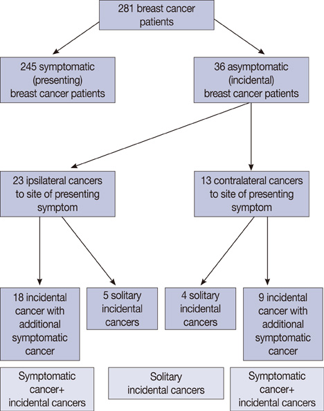
In the study period, 281 women were diagnosed with breast cancer out of 4,400 patients seen at the one-stop breast clinic. Thirty six patients (12.8%) diagnosed with breast cancer had an incidental cancer which was only identified by imaging. The majority of contralateral, incidental cancers were identified by both mammography and ultrasound (US) and patients were all above 35 years.
CONCLUSION
We suggest mammography of both breasts and US of the symptomatic breast in order to identify incidental cancers.
Keyword
Figure
Cited by 1 articles
-
Commentary on: Incidental Breast Cancers Identified in a One-Stop Symptomatic Breast Clinic
Jeong Eon Lee, Jung-Hyun Yang, Seok Jin Nam
J Breast Cancer. 2011;14(2):165-166. doi: 10.4048/jbc.2011.14.2.165.
Reference
-
1. NHS Executive. Improving Outcomes in Breast Cancer: the Manual. 1996. London: NHS Executive.2. Chow WH, Devesa SS, Warren JL, Fraumeni JF Jr. Rising incidence of renal cell cancer in the United States. JAMA. 1999. 281:1628–1631.
Article3. Yoon DY, Chang SK, Choi CS, Yun EJ, Seo YL, Nam ES, et al. The prevalence and significance of incidental thyroid nodules identified on computed tomography. J Comput Assist Tomogr. 2008. 32:810–815.
Article4. Shojaku H, Seto H, Iwai H, Kitazawa S, Fukushima W, Saito K. Detection of incidental breast tumors by noncontrast spiral computed tomography of the chest. Radiat Med. 2008. 26:362–367.
Article5. Hussain A, Gordon-Dixon A, Almusawy H, Sinha P, Desai A. The incidence and outcome of incidental breast lesions detected by computed tomography. Ann R Coll Surg Engl. 2010. 92:124–126.
Article6. NHS Information Centre for Health and Social Care. Breast Screening Programme, England 2007-08. 2009. Leeds: NHS Information Centre for Health and Social Care.7. Simpson WL Jr, Hermann G, Rausch DR, Sherman J, Feig SA, Bleiweiss IJ, et al. Ultrasound detection of nonpalpable mammographically occult malignancy. Can Assoc Radiol J. 2008. 59:70–76.8. Kolb TM, Lichy J, Newhouse JH. Comparison of the performance of screening mammography, physical examination and breast US and evaluation of factors that influence them: an analysis of 27, 825 patient evaluations. Radiology. 2002. 225:165–175.
Article9. Buchberger W, DeKoekkoek-Doll P, Springer P, Obrist P, Dunser M. Incidental findings on sonography of the breast: clinical significance and diagnostic workup. AJR Am J Roentgenol. 1999. 173:921–927.
Article10. Kaplan SS. Clinical utility of bilateral whole-breast US in the evaluation of women with dense breast tissue. Radiology. 2001. 221:641–649.
Article11. Wilkinson LS, Given-Wilson R, Hall T, Potts H, Sharma AK, Smith E. Increasing the diagnosis of multifocal primary breast cancer by the use of bilateral whole-breast ultrasound. Clin Radiol. 2005. 60:573–578.
Article12. Chaudary MA, Millis RR, Hoskins EO, Halder M, Bulbrook RD, Cuzick J, et al. Bilateral primary breast cancer: a prospective study of disease incidence. Br J Surg. 1984. 71:711–714.
Article13. Robbins GF, Berg JW. Bilateral primary breast cancer; a prospective clinicopathological study. Cancer. 1964. 17:1501–1527.
Article14. Roubidoux MA, Helvie MA, Lai NE, Paramagul C. Bilateral breast cancer: early detection with mammography. Radiology. 1995. 196:427–431.
Article15. Muttarak M, Pojchamarnwiputh S, Padungchaichote W, Chaiwun B. Evaluation of the contralateral breast in patients with ipsilateral breast carcinoma: the role of mammography. Singapore Med J. 2002. 43:229–233.16. Berg WA, Gutierrez L, NessAiver MS, Carter WB, Bhargavan M, Lewis RS, et al. Diagnostic accuracy of mammography, clinical examination, US and MR imaging in preoperative assessment of breast cancer. Radiology. 2004. 233:830–849.
Article17. Berg WA, Gilbreath PL. Multicentric and multifocal cancer: whole-breast US in preoperative evaluation. Radiology. 2000. 214:59–66.
Article18. Biggs MJ, Ravichandran D. Mammography in symptomatic women attending a rapid diagnosis breast clinic: a prospective study. Ann R Coll Surg Engl. 2006. 88:306–308.
Article19. Sterns EE. The abnormal mammogram in women with clinically normal breasts. Can J Surg. 1995. 38:168–172.20. Kerin MJ, O'Hanlon DM, Khalid AA, Kent PJ, McCarthy PA, Given HF. Mammographic assessment of the symptomatic nonsuspicious breast. Am J Surg. 1997. 173:181–184.
Article
- Full Text Links
- Actions
-
Cited
- CITED
-
- Close
- Share
- Similar articles
-
- Commentary on: Incidental Breast Cancers Identified in a One-Stop Symptomatic Breast Clinic
- Incidental Extramammary Findings on Preoperative Breast MRI in Breast Cancer Patients: A Pictorial Essay
- Current State and Role of Private Breast Clinic in Korea
- Bilateral breast carcinoma
- Breast Cancer Screening of 13,791 Women by Physical Examination and Mammography


