Chordoid Glioma Originating in the Intrasellar and Suprasellar Regions: Case Report
- Affiliations
-
- 1Department of Radiology, Soonchunhyang University Bucheon Hospital, Bucheon, Korea. aleerad@schmc.ac.kr
- 2Department of Pathology, Soonchunhyang University Bucheon Hospital, Bucheon, Korea.
- 3Department of Neurosurgery, Soonchunhyang University Bucheon Hospital, Bucheon, Korea.
- KMID: 2175592
- DOI: http://doi.org/10.13104/imri.2015.19.2.117
Abstract
- Chordoid glioma is a rare, low-grade brain neoplasm typically located in the third ventricle. Herein, we report an unusual case of histologically confirmed chordoid glioma located in the pituitary fossa and suprasellar region, not attached to the third ventricle. A 57-year-old woman presented with a 2-month history of headache and visual disturbance. Magnetic resonance imaging revealed an ovoid mass in the pituitary fossa and suprasellar region, compressing the optic chiasm without involvement of the third ventricle. The tumor showed low signal intensity on T1-weighted images and iso- to high signal intensity on T2-weighted images, with strong and homogenous contrast enhancement. Subtotal resection was performed via the transcranial approach, and the patient subsequently received adjuvant gamma knife radiosurgery. However, the residual mass showed disease progression 5 months after the initial surgery.
MeSH Terms
Figure
-
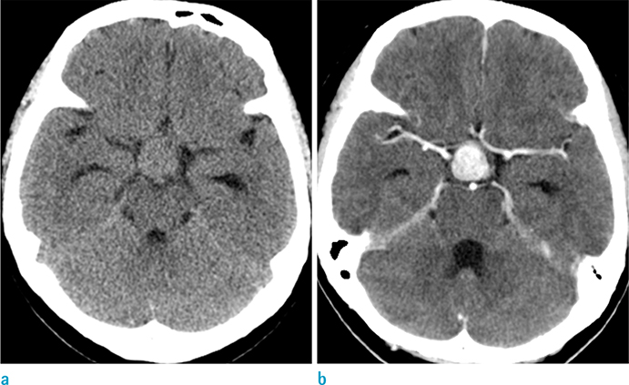
Fig. 1 An axial precontrast (a) and a contrast-enhanced (b) computed tomography scan show an oval, well-defined, and hyperdense mass with strong and homogenous enhancement in the sellar and suprasellar regions.
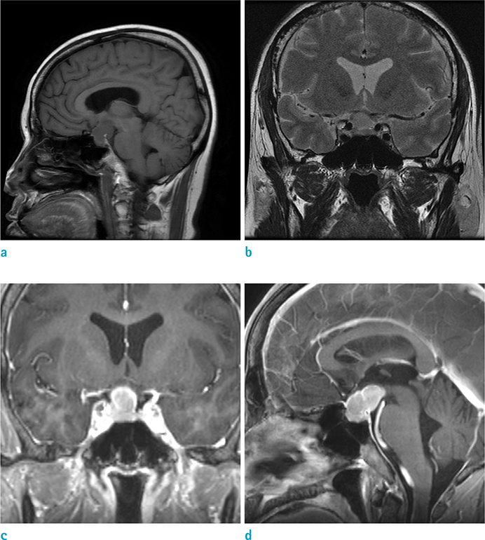
Fig. 2 Magnetic resonance images of the tumor. (a) Sagittal T1-weighted image shows low signal intensity of the mass. (b) Coronal T2-weighted image shows iso- to high signal intensity of the mass. Coronal (c) and sagittal (d) contrast-enhanced T1-weighted images of the brain reveal a strong, homogeneously enhancing solid sellar and suprasellar mass, not connected to the third ventricle. The normal pituitary gland and stalk are not discernible. The epicenter of the mass appears to originate in the sella, and not in the lamina terminalis of the anterior third ventricle.

Fig. 3 Histological features of the tumor. (a) The tumor shows clusters and cords of epithelioid-to-spindle cells embedded in a myxoid matrix and inflammatory cells (Hematoxylin & Eosin staining, original magnification × 100). (b) The positive immunoreactivity for glial fibrillary acidic protein expression are visualized as brown (original magnification × 200).
Reference
-
1. Brat DJ, Scheithauer BW, Staugaitis SM, Cortez SC, Brecher K, Burger PC. Third ventricular chordoid glioma: a distinct clinicopathologic entity. J Neuropathol Exp Neurol. 1998; 57:283–290.2. Pomper MG, Passe TJ, Burger PC, Scheithauer BW, Brat DJ. Chordoid glioma: a neoplasm unique to the hypothalamus and anterior third ventricle. AJNR Am J Neuroradiol. 2001; 22:464–469.3. Kim JW, Kim JH, Choe G, Kim CY. Chordoid glioma: a case report of unusual location and neuroradiological characteristics. J Korean Neurosurg Soc. 2010; 48:62–65.4. Suh YL, Kim NR, Kim JH, Park SH. Suprasellar chordoid glioma combined with Rathke's cleft cyst. Pathol Int. 2003; 53:780–785.5. Pasquier B, Peoc'h M, Morrison AL, et al. Chordoid glioma of the third ventricle: a report of two new cases, with further evidence supporting an ependymal differentiation, and review of the literature. Am J Surg Pathol. 2002; 26:1330–1342.6. Grand S, Pasquier B, Gay E, Kremer S, Remy C, Le Bas JF. Chordoid glioma of the third ventricle: CT and MRI, including perfusion data. Neuroradiology. 2002; 44:842–846.7. Al Hinai QS, Petrecca K. Rarest of the rare: chordoid glioma infiltrating the optic chiasm. Surg Neurol Int. 2011; 2:53.8. Cenacchi G, Roncaroli F, Cerasoli S, Ficarra G, Merli GA, Giangaspero F. Chordoid glioma of the third ventricle: an ultrastructural study of three cases with a histogenetic hypothesis. Am J Surg Pathol. 2001; 25:401–405.9. Donovan JL, Nesbit GM. Distinction of masses involving the sella and suprasellar space: specificity of imaging features. AJR Am J Roentgenol. 1996; 167:597–603.10. Kobayashi T, Tsugawa T, Hashizume C, et al. Therapeutic approach to chordoid glioma of the third ventricle. Neurol Med Chir (Tokyo). 2013; 53:249–225.
- Full Text Links
-
- Actions
-
Cited
- CITED
-
- Close
- Share
-
- Similar articles
-
- Suprasellar Chordoid Glioma Combined with Rathke's Cleft Cyst: Case Report
- Chordoid Glioma: an Uncommon Tumor of the Third Ventricle
- Chordoid Glioma of the Third Ventricle with Unusual MRI Features
- Chordoid Glioma in the Third Ventricle: Case Report
- Chordoid Glioma with Intraventricular Dissemination: A Case Report with Perfusion MR Imaging Features
