Neuroimaging and Clinicopathologic Findings of Lymphoplasmacyte-rich Meningioma, Mimicking Malignancy: Case Report
- Affiliations
-
- 1Department of Radiology, Seoul St. Mary's Hospital, The Catholic University of Korea, Seoul, Korea. ahn-kj@catholic.ac.kr
- 2Department of Hospital Pathology, Seoul St. Mary's Hospital, The Catholic University of Korea, Seoul, Korea.
- 3Department of Neurosurgery, Seoul St. Mary's Hospital, The Catholic University of Korea, Seoul, Korea.
- KMID: 2175579
- DOI: http://doi.org/10.13104/imri.2015.19.1.62
Abstract
- Lymphoplasmacyte-rich meningioma is a rare WHO Grade I subtype of meningioma. The lymphoplasmacyte-rich meningioma does not have typical imaging features of a meningioma so it can mimic intracranial inflammatory condition or brain neoplasm. We report the clinicopathologic features of lymphoplasmacyte-rich meningioma in a 35-year-old woman. She suffered from progressive headache, dizziness and tinnitus over two years. The tumor exhibited atypical neuroimaging features, including obvious peritumoral edema and irregular enhancing components. She underwent total resection and histologic examination revealed a meningioma with numerous plasma cells. Her symptoms have since resolved and there has been no evidence of tumor recurrence after one year of follow-up.
MeSH Terms
Figure
-
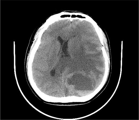
Fig. 1 Non-enhanced CT demonstrates a cystic tumor mass with an internal fluid-fluid level and extensive brain edema, resulting in mid-line shift.

Fig. 2 The lesion demonstrates cystic component with hemorrhagic fluid-fluid level and extensive edematous change on axial T2-weighted image (a) and axial T1-weighted image (b). Irregular thick enhancing wall is noted on contrast-enhanced T1-weighted axial image (c). The mass shows broad dural based attachment on contrast-enhanced T1-weighted sagittal image (d).
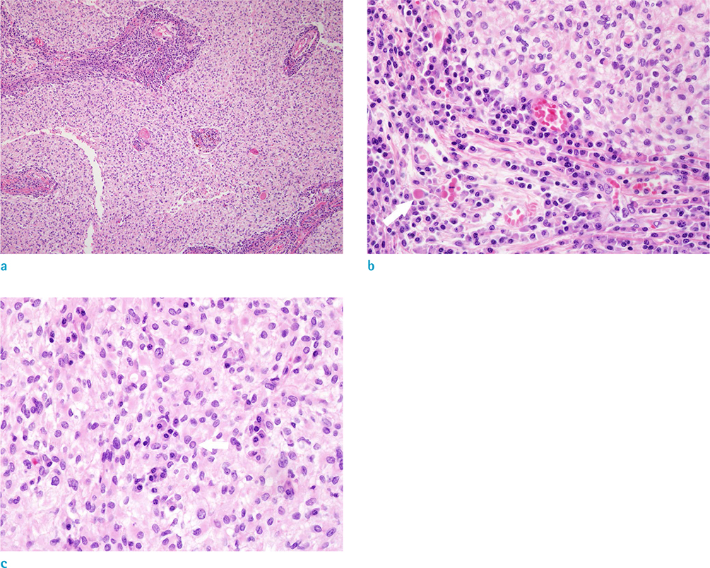
Fig. 3 Photomicrograph (H & E staining) with low magnification view (a) shows lobules with massive plasma cell infiltration. High magnification view (b) demonstrates numerous plasma cells with Russel bodies (arrow). The tumor cells shows a few nuclear pseudoinclusions (c)
Reference
-
1. Marosi C, Hassler M, Roessler K, et al. Meningioma. Crit Rev Oncol Hematol. 2008; 67:153–171.2. Louis DN, Ohgaki H, Weistler OD, et al. The 2007 WHO classification of tumours of the central nervous system. Acta Neuropathol. 2007; 114:97–109.3. Liu Jl, Zhou Jl, Ma YH, Dong C. An analysis of the magnetic resonance imaging and pathology of intracal lymphoplasmacyte-rich meningioma. Eur J Radiol. 2012; 81:968–973.4. Zhu HD, Xie Q, Gong Y, et al. Lymphoplasmacyte-rich meningioma: our experience with 19 cases and a systematic literature reviews. Int J Clin Exp Med. 2013; 6:504–515.5. Yamaki T, Ikeda T, Sakamoto Y, Ohtaki M, Hashi K. Lymphoplasmacyte-rich meningioma with clinical resemblance to inflammatory pseudotumor. Report two cases. J Neurosurg. 1997; 86:898–904.6. Lee KJ, Joo WI, Rha HK, et al. Peritumoral brain edema in meningiomas: correlations between magnetic resonance imaging, angiography, and pathology. Surg Neurol. 2008; 69:350–355.7. Banerjee AK, Blackwood W. A subfrontal tumor with the features of plasmocytoma and meningioma. Acta Neuropathol. 1971; 18:84–88.8. Bruno MC, Ginguené C, Santangelo M, et al. Lymphoplasacyte rich meningioma. A case report and review of the literature. J Neurosurg Sci. 2004; 48:117–124.9. Horten BC, Urich H, Stefoski D. Meningiomas with conspicuous plasma cell-lymphocystic components: a reportion of five cases. Cancer. 1979; 43:258–264.10. Loh JK, Hwang SL, Tsai KB, Kwan AL, Howng SL. Sphenoid ridge lymphoplasmacyte-rich meningioma. J Formos Med Assoc. 2006; 105:594–598.
- Full Text Links
-
- Actions
-
Cited
- CITED
-
- Close
- Share
-
- Similar articles
-
- Extradural Spinal Lymphoplasmacyte-Rich Meningioma in the Thoracic Spine: A Case Report and Literature Review
- Primary Extracranial Meningioma Mimicking Musculoskeletal Malignancy: A Case Report
- Ewing's Sarcoma Mimicking a Meningioma in Radiological Findings: A Case Report
- Primary Pulmonary Meningioma Mimicking Lung Metastasis: A Case Report
- Primary Intraosseous Meningioma
