Ann Dermatol.
2015 Aug;27(4):467-468. 10.5021/ad.2015.27.4.467.
A Case of Sporadic Dyschromatosis Universalis Hereditaria
- Affiliations
-
- 1Department of Dermatology, Soonchunhyang University College of Medicine, Seoul, Korea. snolomas@schmc.ac.kr
- KMID: 2171514
- DOI: http://doi.org/10.5021/ad.2015.27.4.467
Abstract
- No abstract available.
Figure
-
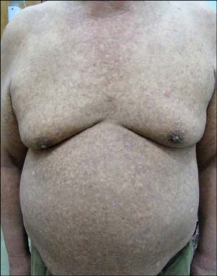
Fig. 1 Generalized reticulated hyper- and hypo-pigmented macules with scales on the whole body.
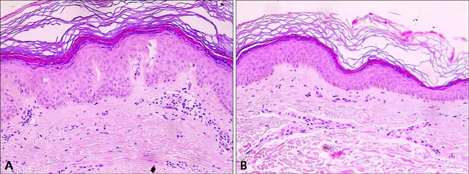
Fig. 2 Skin biopsies were collected from hyper- and hypopigmented lesions on the upper arm (H&E; ×200). (A) Marked decreased basal pigmentation in a hypopigmented lesion. (B) Slightly increased melanin content in the basal layer of a hyperpigmented lesion.
Reference
-
1. Bukhari IA, El-Harith EA, Stuhrmann M. Dyschromatosis universalis hereditaria as an autosomal recessive disease in five members of one family. J Eur Acad Dermatol Venereol. 2006; 20:628–629.
Article2. Sethuraman G, Srinivas CR, D'Souza M, Thappa DM, Smiles L. Dyschromatosis universalis hereditaria. Clin Exp Dermatol. 2002; 27:477–479.
Article3. Kim NS, Im S, Kim SC. Dyschromatosis universalis hereditaria: an electron microscopic examination. J Dermatol. 1997; 24:161–164.
Article4. Liu H, Li Y, Hung KK, Wang N, Wang C, Chen X, et al. Genome-wide linkage, exome sequencing and functional analyses identify ABCB6 as the pathogenic gene of dyschromatosis universalis hereditaria. PLoS One. 2014; 9:e87250.
Article5. Ro YS, Nam TS, Lee CW, Park CK, Seou WP, Kim JH. Dyschromatosis universalis hereditaria. Ann Dermatol. 1990; 2:24–30.
Article
