Ewha Med J.
2016 Apr;39(2):45-50. 10.12771/emj.2016.39.2.45.
Microarray Analysis in Spontaneously Hypertensive Rat Heart after Losartan Treatment
- Affiliations
-
- 1Department of Pediatrics, Ewha Womans University School of Medicine, Seoul, Korea. ymhong@ewha.ac.kr
- 2Department of Thoracic and Cardiovascular Surgery, Ewha Womans University School of Medicine, Seoul, Korea.
- KMID: 2171364
- DOI: http://doi.org/10.12771/emj.2016.39.2.45
Abstract
OBJECTIVES
Spontaneously hypertensive rats (SHR) are frequently used as rat models of essential hypertension. The mechanism for the development of hypertension is complicated and it is unknown. The renin-angiotensin system (RAS) plays a key role in the control of blood pressure. Microarrays are a powerful tool for studying genetics. The purpose of this study was to investigate changes of gene expression in the heart tissues of SHR after losartan treatment to provide basic data that is useful in the early diagnosis of hypertension and gene treatment.
METHODS
Rats were divided into three groups: the control (C) group; the hypertension (H) group (SHR), and the losartan (L) group; treated with losartan (10 mg/kg/day) in SHR. Rats were sacrificed at week 5 and microarray analysis was performed.
RESULTS
102 gene expressions including the genes associated with cell proliferation such as Raf1, Uchl1, Btla, Spock1 were increased. The other 139 gene expressions, including the genes related to the regulation of metabolism such as TFIID, Auf1, Bmp, Hub, Taf51 showed decreases in gene expression. A total of 31 genes were differentially expressed in the L group compared to the H group. Of these, 16 genes including the genes associated with macromolecule metabolism such as MGC105766, Ppp1r1a, Rpl3l showed increased expression. The other 15 genes including the genes associated with primary metabolism such as Mcpt4, Ngn3, Tdo, Ak2 Hyal2 showed decreased expressions.
CONCLUSION
According to microarray analysis, there was significant gene expression change in SHR compared with normal rats as well as significant gene expression changes after losartan treatment in SHR.
Keyword
MeSH Terms
Figure
-
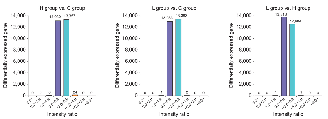
Fig. 1 Intensity ratio histograph in three groups. The graph shows a number of genes showing gene expression changes. C, control; H, hypertension; L, losartan.
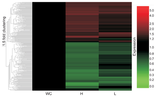
Fig. 2 Changes of gene heat map after losartan treatment. This is the gene expression profile expressed differentially over 1.5 fold (P<0.05). Red color represents over expression and green color represents down expression. C, control; H, hypertension; L, losartan.
Reference
-
1. Delles C, McBride MW, Graham D, Padmanabhan S, Dominiczak AF. Genetics of hypertension: from experimental animals to humans. Biochim Biophys Acta. 2010; 1802:1299–1308.2. Choi SM, Seo MJ, Kang KK, Kim JH, Ahn BO, Yoo M. Beneficial effects of the combination of amlodipine and losartan for lowering blood pressure in spontaneously hypertensive rats. Arch Pharm Res. 2009; 32:353–358.3. Okamoto K, Aoki K. Development of a strain of spontaneously hypertensive rats. Jpn Circ J. 1963; 27:282–293.4. Zhang H, Wu S, Huang C, Li X. Long term treatment of spontaneously hypertensive rats with losartan and molecular basis of modulating Ito of ventricular myocytes. Mol Med Rep. 2014; 9:1959–1967.5. Ravassa S, Fortuno MA, Gonzalez A, Lopez B, Zalba G, Fortuno A, et al. Mechanisms of increased susceptibility to angiotensin II-induced apoptosis in ventricular cardiomyocytes of spontaneously hypertensive rats. Hypertension. 2000; 36:1065–1071.6. Cha JH, Lee HR, Kim KC, Cho MS, Hong YM. Changes of gene expressions in spontaneously hypertensive rat model after losartan treatment. Korean Circ J. 2012; 42:761–768.7. Dai Q, Xu M, Yao M, Sun B. Angiotensin AT1 receptor antagonists exert anti-inflammatory effects in spontaneously hypertensive rats. Br J Pharmacol. 2007; 152:1042–1048.8. Diep QN, El Mabrouk M, Yue P, Schiffrin EL. Effect of AT(1) receptor blockade on cardiac apoptosis in angiotensin II-induced hypertension. Am J Physiol Heart Circ Physiol. 2002; 282:H1635–H1641.9. Kobori H, Nangaku M, Navar LG, Nishiyama A. The intrarenal renin-angiotensin system: from physiology to the pathobiology of hypertension and kidney disease. Pharmacol Rev. 2007; 59:251–287.10. Nishiyama A, Hitomi H, Rahman A, Kiyomoto H. Drug discovery for overcoming chronic kidney disease (CKD): pharmacological effects of mineralocorticoid-receptor blockers. J Pharmacol Sci. 2009; 109:1–6.11. Cagnoni F, Njwe CA, Zaninelli A, Ricci AR, Daffra D, D'Ospina A, et al. Blocking the RAAS at different levels: an update on the use of the direct renin inhibitors alone and in combination. Vasc Health Risk Manag. 2010; 6:549–559.12. Koprdova R, Cebova M, Kristek F. Long-term effect of losartan administration on blood pressure, heart and structure of coronary artery of young spontaneously hypertensive rats. Physiol Res. 2009; 58:327–335.13. Buse JB, Ginsberg HN, Bakris GL, Clark NG, Costa F, Eckel R, et al. Primary prevention of cardiovascular diseases in people with diabetes mellitus: a scientific statement from the American Heart Association and the American Diabetes Association. Circulation. 2007; 115:114–126.14. Mancia G, De Backer G, Dominiczak A, Cifkova R, Fagard R, Germano G, et al. 2007 Guidelines for the Management of Arterial Hypertension: The Task Force for the Management of Arterial Hypertension of the European Society of Hypertension (ESH) and of the European Society of Cardiology (ESC). J Hypertens. 2007; 25:1105–1187.15. Fortuno MA, Ravassa S, Etayo JC, Diez J. Overexpression of Bax protein and enhanced apoptosis in the left ventricle of spontaneously hypertensive rats: effects of AT1 blockade with losartan. Hypertension. 1998; 32:280–286.16. Zhao LL, Chen HJ, Chen JZ, Yu M, Ni YL, Zhang WF. Losartan reduced connexin43 expression in left ventricular myocardium of spontaneously hypertensive rats. J Zhejiang Univ Sci B. 2008; 9:448–454.17. Yamamoto H, Okuzaki D, Yamanishi K, Xu Y, Watanabe Y, Yoshida M, et al. Genetic analysis of genes causing hypertension and stroke in spontaneously hypertensive rats. Int J Mol Med. 2013; 31:1057–1065.18. McBride MW, Charchar FJ, Graham D, Miller WH, Strahorn P, Carr FJ, et al. Functional genomics in rodent models of hypertension. J Physiol. 2004; 554:56–63.19. Okamoto K, Hazama F, Yamori Y, Haebara H, Nagaoka A. Pathogenesis and prevention of stroke in spontaneously hypertensive rats. Clin Sci Mol Med Suppl. 1975; 2:161s–163s.20. Qin XP, Zeng SY, Tian HH, Deng SX, Ren JF, Zheng YB, et al. Involvement of prolylcarboxypeptidase in the effect of rutaecarpine on the regression of mesenteric artery hypertrophy in renovascular hypertensive rats. Clin Exp Pharmacol Physiol. 2009; 36:319–324.21. Akioyamen L, Levine M, Sherifali D, O'Reilly D, Frankfurter C, Pullenayegum E, et al. Cardiovascular and cerebrovascular outcomes of long-term angiotensin receptor blockade: meta-analyses of trials in essential hypertension. J Am Soc Hypertens. 2016; 10:55–69.e1.22. ESH/ESC Task Force for the Management of Arterial Hypertension. 2013 Practice guidelines for the management of arterial hypertension of the European Society of Hypertension (ESH) and the European Society of Cardiology (ESC): ESH/ESC Task Force for the Management of Arterial Hypertension. J Hypertens. 2013; 31:1925–1938.23. Zhao Q, Kelly TN, Li C, He J. Progress and future aspects in genetics of human hypertension. Curr Hypertens Rep. 2013; 15:676–686.
- Full Text Links
-
- Actions
-
Cited
- CITED
-
- Close
- Share
-
- Similar articles
-
- Changes of Bax, Bcl-2, CCR-2, MCP-1, and TGF-β1 genes in the left ventricle of spontaneously hypertensive rat after losartan treatment
- Changes of Gene Expressions in Spontaneously Hypertensive Rat Model After Losartan Treatment
- Effects of Centrally Administered Losartan on Deoxycorticosterone-salt Hypertension Rats
- Effect of Central Losartan on DOCA-Salt Hypertension Rats
- Comparison of ANG II-mediated [Ca2+i, IP3 Production and [ATP]i in Isolated Renal Proximal Convoluted Tubules of Adult SHR and WKY
