Gastric Emphysema Related with Superior Mesenteric Artery Syndrome
- Affiliations
-
- 1Department of Internal Medicine, Jeju National University School of Medicine, Jeju, Korea. songhj@jejunu.ac.kr
- 2Department of Radiology, Jeju National University School of Medicine, Jeju, Korea.
- KMID: 2171299
- DOI: http://doi.org/10.12771/emj.2014.37.2.141
Abstract
- Gastric emphysema is caused by a mucosal disruption of stomach, which is leading to the dissection of air into the wall. A 24-year-old man admitted to our hospital with vomiting, abdominal distension, and pain. Abdominal computed tomography showed severe gastric distension, air within the gastric wall, and a compressed third segment of the duodenum by superior mesenteric artery (SMA). The upper endoscopy revealed multiple geographic ulcers in the gastric body and marked dilatation of the second segment of duodenum and a collapsed third segment. Based on these findings and his symptoms, the patient was diagnosed as having gastric emphysema related with SMA syndrome. He improved after the nasogastric decompression, jejunal feeding and administration of antibiotics. We report a rare case of gastric emphysema related with SMA syndrome. He was managed successfully with medical treatment and nutritional support.
MeSH Terms
Figure
-

Fig. 1 Contrast enhanced abdominal computed tomography scan. It shows severe gastric distention with air-fluid level and fluid-filled duodenal dilatation. Extrinsic compression of third segment of duodenum between superior mesenteric artery (SMA) and abdominal aorta and a narrow aortomesenteric angle to 12° (A, black arrows) are suggestive of SMA syndrome. Multiple thin linear air densities within the distended gastric wall is suggestive of gastric emphysema (B, white arrow).
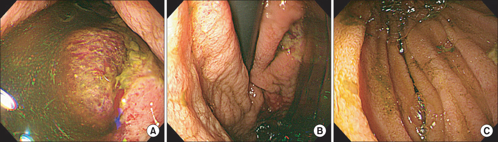
Fig. 2 Upper endoscopy. It shows many geographic gastric ulcers with mucosal hemorrhage in the body of stomach (A, B), and collapsed third segment associated with the superior mesenteric artery syndrome (C).
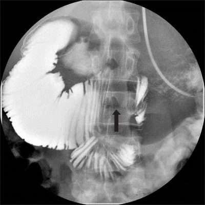
Fig. 3 Nasojejunal feeding tube insertion with fluoroscopic guidance on the fourth day of hospital. Extrinsic compression of third segment of duodenum is compatible with superior mesenteric artery syndrome (black arrow).
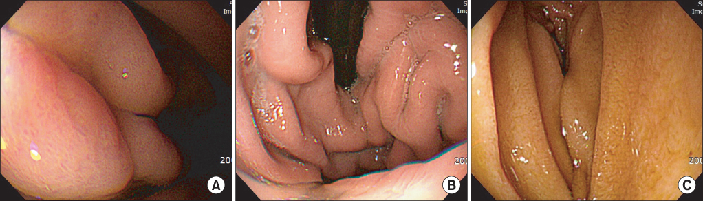
Fig. 4 Follow-up upper endoscopy after 2 months showed complete recovery from the gastric emphysema (A, B) and a mildly constricted third segment of the duodenum by extrinsic superior mesenteric artery compression (C).
Reference
-
1. Soon MS, Yen HH, Soon A, Lin OS. Endoscopic ultrasonographic appearance of gastric emphysema. World J Gastroenterol. 2005; 11:1719–1721.2. Feczko PJ, Mezwa DG, Farah MC, White BD. Clinical significance of pneumatosis of the bowel wall. Radiographics. 1992; 12:1069–1078.3. Klipfel AA, Kessler E, Schein M. Rapunzel syndrome causing gastric emphysema and small bowel obstruction. Surgery. 2003; 133:120–121.4. Fierst SM, Robinson HM, Lasagna L. Interstitial gastric emphysema following gastroscopy: its relation to the syndrome of pneumoperitoneum and generalized emphysema with no evident perforation. Ann Intern Med. 1951; 34:1202–1212.5. Allan K, Barriga J, Afshani M, Davila R, Tombazzi C. Emphysematous gastritis. Am J Med Sci. 2005; 329:205–207.6. Yalamanchili M, Cady W. Emphysematous gastritis in a hemodialysis patient. South Med J. 2003; 96:84–88.7. Hadas-Halpren I, Hiller R, Guberman D. Emphysematous gastritis secondary to ingestion of large amounts of Coca-Cola. Am J Gastroenterol. 1993; 88:127–129.8. Kim EB, Lee TH. Superior mesenteric artery syndrome: past and present. Korean J Med. 2013; 84:28–36.9. Yang HW, Byeon JS, Lee GH, Yang SG, Jung HY, Kim JH, et al. A case of gastric emphysema with portal vein emphysema associated with the episode of severe vomiting. Korean J Gastrointest Endosc. 2005; 31:107–110.10. Moon SW, Lee SW, Choi SH, Hong YS. Gastric emphysema after methyl ethyl ketone peroxide ingestion. Clin Toxicol (Phila). 2010; 48:90–91.11. Hyun YS, Han DS, Lee HL, Bae JH, Eun CS. Gastric emphysema after endoscopic submucosal dissection. Endoscopy. 2011; 43:Suppl 2 UCTN. E83–E84.12. Jeong IB, Kim S, Kim Y. Gastric wall emphysema after endoscopic submucosal tumor biopsy. Clin Gastroenterol Hepatol. 2011; 9:e101–e102.13. Kim TY, Kim HU, Song HJ. A case of gastric emphysema in anorexia nervosa presenting as acute gastric distension. Korean J Gastroenterol. 2012; 60:315–319.14. Kim M, You JR, Kim HU. A case of gastric emphysema associated with superior mesenteric artery syndrome. Korean J Helicobacter Up Gastrointest Res. 2012; 12:120–123.15. van Mook WN, van der Geest S, Goessens ML, Schoon EJ, Ramsay G. Gas within the wall of the stomach due to emphysematous gastritis: case report and review. Eur J Gastroenterol Hepatol. 2002; 14:1155–1160.16. Kussin SZ, Henry C, Navarro C, Stenson W, Clain DJ. Gas within the wall of the stomach report of a case and review of the literature. Dig Dis Sci. 1982; 27:949–954.17. Fishman EK, Urban BA, Hruban RH. CT of the stomach: spectrum of disease. Radiographics. 1996; 16:1035–1054.18. Grayson DE, Abbott RM, Levy AD, Sherman PM. Emphysematous infections of the abdomen and pelvis: a pictorial review. Radiographics. 2002; 22:543–561.19. Cordum NR, Dixon A, Campbell DR. Gastroduodenal pneumatosis: endoscopic and histological findings. Am J Gastroenterol. 1997; 92:692–695.
- Full Text Links
-
- Actions
-
Cited
- CITED
-
- Close
- Share
-
- Similar articles
-
- A Case of Gastric Emphysema Associated with Superior Mesenteric Artery Syndrome
- A Case of Gastric Emphysema in Anorexia Nervosa Presenting as Acute Gastric Distension
- Case Report: Superior Mesenteric Artery Syndrome following Laparoscopic Adjustable Gastric Banding
- Superior mesenteric artery syndrome with achalasia
- A case of the superior mesenteric artery syndrome
