Effects of Serum 25-hydroxy-vitamin D and Fetal Bone Growth during Pregnancy
- Affiliations
-
- 1Division of Endocrinoloy, Cheil General Hospital & Women's Healthcare Center, Dankook University College of Medicine, Seoul, Korea. hyunkooyoon@hotmail.com
- 2Department of Obstetrics & Gynecology, Cheil General Hospital & Women's Healthcare Center, Dankook University College of Medicine, Seoul, Korea.
- KMID: 2170109
- DOI: http://doi.org/10.11005/jbm.2015.22.3.127
Abstract
- BACKGROUND
This study was conducted to observe the prevalence of vitamin D deficiency during pregnancy and the effects of maternal 25-hydroxy-vitamin D (25-[OH]D) levels on fetal bone growth.
METHODS
Five hundred twenty-three Korean pregnant women were randomly recruited and serum 25-(OH)D level was measured. During pregnancy, fetal ultrasonography and serum 25-(OH)D measurements were carried out 3 times in 275 of 523 pregnant women. Fetal biparietal and occipitofrontal diameter, head and abdominal circumference, and femur and humerus length were measured through fetal ultrasonography.
RESULTS
The prevalence of vitamin D deficiency (25-[OH]D<20 ng/mL) based on the 1st serum measurement of 25-(OH)D was 88.9%. There was no association between maternal serum 25-(OH)D level and fetal bone growth. In 275 pregnant women who completed study design, the mean value of 25-(OH)D was 12.97+/-5.93, 19.12+/-9.82, and 19.60+/-9.98 ng/mL at 12 to 14, 20 to 22, and 32 to 34 weeks of pregnancy, respectively and there was an association between the difference of serum 25-(OH)D level between 12 to 14 and 20 to 22 weeks and growth velocity of fetal biparietal diameter between 20 to 22 and 32 to 34 weeks of pregnancy.
CONCLUSIONS
This study shows a high prevalence of vitamin D deficiency in Korean pregnant women and the change of serum 25-(OH)D levels is related with the growth of fetal biparietal diameter, however other parameters are not associated with serum 25-(OH)D levels.
Keyword
MeSH Terms
Figure
-
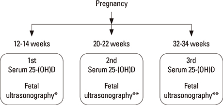
Fig. 1 Flow of measurements for participants in this study. 275 among 523 early pregnant women were completed the blood samplings and ultrasonographys three times. 25-(OH)D, 25-hydroxy-vitamin D. Parameters for measurement. *biparietal diameter (BPD), abdominal circumference (AC); **biparietal diameter (BPD), occipitofrontal diameter (OFD), head circumference (HC), abdominal circumference (AC) femur length (FL), humerus length (HL).

Fig. 2 Comparison of fetal growth velocity between pregnant women whose serum 25-(OH)D level was higher than 20 ng/mL and those who were 25-(OH)D deficient. (A) No association between serum 25-(OH)D levels at 12 to 14 weeks and the growth velocity between 12 to 14 and 20 to 22 weeks of pregnancy was observed. (B) The growth velocity between 12 to 14 and 20 to 22 weeks and between 20 to 22 and 32 to 34 weeks of pregnancy were not associated with serum 25-(OH)D levels at 20 to 22 weeks. (C) No association between serum 25-(OH)D levels at 32 to 34 weeks and the growth velocity between 20 to 22 and 32 to 34 weeks of pregnancy was observed. 25-(OH)D, 25-hydroxy-vitamin D; BPD, biparietal diameter; AC, abdominal circumference; OFD, occipitofrontal diameter; HC, head circumference; FL, femur length; HL, humerus length. BPD (1), AC(1) : growth velocity between 12-14 and 20-22 weeks of pregnancy. BPD(2), AC(2), OFD(2), HC(2), FL(2), HL(2): growth velocity between 20-22 and 32-34 weeks of pregnancy.
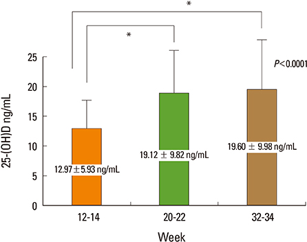
Fig. 3 Mean value of serum 25-(OH)D at 12 to 14, 20 to 22, 32 to 34 weeks of pregnancy in 275 pregnant women who completed study design. Mean value of serum 25-(OH)D at 12 to 14 weeks was significantly different from the mean value at 20 to 22 and 32 to 34 weeks of pregnancy. 25-(OH)D, 25-hydroxy-vitamin D.

Fig. 4 Correlation of the difference of serum 25-(OH)D levels between 12 to 14 and 20 to 22 weeks with the growth velocity of fetal biparietal diameter between 20 to 22 and 32 to 34 weeks of pregnancy in 275 pregnant women who completed study design. A positive correlation was observed between the difference of serum 25-(OH)D levels between 12 to 14 and 20 to 22 weeks and growth velocity of fetal biparietal diameter between 20 to 22 and 32 to 34 weeks of pregnancy. 25-(OH)D, 25-hydroxy-vitamin D.
Cited by 1 articles
-
Gestational Diabetes Mellitus, Fetal Growth and Vitamin D
Hyun Koo Yoon
J Bone Metab. 2017;24(3):155-159. doi: 10.11005/jbm.2017.24.3.155.
Reference
-
1. Holick MF. The vitamin D deficiency pandemic and consequences for nonskeletal health: mechanisms of action. Mol Aspects Med. 2008; 29:361–368.
Article2. Bikle D. Nonclassic actions of vitamin D. J Clin Endocrinol Metab. 2009; 94:26–34.
Article3. Brannon PM, Picciano MF. Vitamin D in pregnancy and lactation in humans. Annu Rev Nutr. 2011; 31:89–115.
Article4. Lewis S, Lucas RM, Halliday J, et al. Vitamin D deficiency and pregnancy: from preconception to birth. Mol Nutr Food Res. 2010; 54:1092–1102.
Article5. Lapillonne A. Vitamin D deficiency during pregnancy may impair maternal and fetal outcomes. Med Hypotheses. 2010; 74:71–75.
Article6. Bodnar LM, Catov JM, Simhan HN, et al. Maternal vitamin D deficiency increases the risk of preeclampsia. J Clin Endocrinol Metab. 2007; 92:3517–3522.
Article7. Morley R, Carlin JB, Pasco JA, et al. Maternal 25-hydroxyvitamin D and parathyroid hormone concentrations and offspring birth size. J Clin Endocrinol Metab. 2006; 91:906–912.
Article8. Merewood A, Mehta SD, Chen TC, et al. Association between vitamin D deficiency and primary cesarean section. J Clin Endocrinol Metab. 2009; 94:940–945.
Article9. Dawodu A, Akinbi H. Vitamin D nutrition in pregnancy: current opinion. Int J Womens Health. 2013; 5:333–343.
Article10. Mulligan ML, Felton SK, Riek AE, et al. Implications of vitamin D deficiency in pregnancy and lactation. Am J Obstet Gynecol. 2010; 202:429.e1–429.e9.
Article11. Dror DK, Allen LH. Vitamin D inadequacy in pregnancy: biology, outcomes, and interventions. Nutr Rev. 2010; 68:465–477.
Article12. Wagner CL, Taylor SN, Dawodu A, et al. Vitamin D and its role during pregnancy in attaining optimal health of mother and fetus. Nutrients. 2012; 4:208–230.
Article13. Holick MF, Binkley NC, Bischoff-Ferrari HA, et al. Evaluation, treatment, and prevention of vitamin D deficiency: an Endocrine Society clinical practice guideline. J Clin Endocrinol Metab. 2011; 96:1911–1930.
Article14. Galthen-Sørensen M, Andersen LB, Sperling L, et al. Maternal 25-hydroxyvitamin D level and fetal bone growth assessed by ultrasound: a systematic review. Ultrasound Obstet Gynecol. 2014; 44:633–640.
Article15. Cross NA, Hillman LS, Allen SH, et al. Calcium homeostasis and bone metabolism during pregnancy, lactation, and postweaning: a longitudinal study. Am J Clin Nutr. 1995; 61:514–523.
Article16. Zhang JY, Lucey AJ, Horgan R, et al. Impact of pregnancy on vitamin D status: a longitudinal study. Br J Nutr. 2014; 112:1081–1087.
Article17. Bouillon R, Van Assche FA, Van Baelen H, et al. Influence of the vitamin D-binding protein on the serum concentration of 1,25-dihydroxyvitamin D3. Significance of the free 1,25-dihydroxyvitamin D3 concentration. J Clin Invest. 1981; 67:589–596.
Article18. Novakovic B, Sibson M, Ng HK, et al. Placenta-specific methylation of the vitamin D 24-hydroxylase gene: implications for feedback autoregulation of active vitamin D levels at the fetomaternal interface. J Biol Chem. 2009; 284:14838–14848.19. Powe CE, Evans MK, Wenger J, et al. Vitamin D-binding protein and vitamin D status of black Americans and white Americans. N Engl J Med. 2013; 369:1991–2000.
Article20. Viljakainen HT, Saarnio E, Hytinantti T, et al. Maternal vitamin D status determines bone variables in the newborn. J Clin Endocrinol Metab. 2010; 95:1749–1757.
Article21. Mahon P, Harvey N, Crozier S, et al. Low maternal vitamin D status and fetal bone development: cohort study. J Bone Miner Res. 2010; 25:14–19.
Article22. Hamilton SA, McNeil R, Hollis BW, et al. Profound Vitamin D Deficiency in a Diverse Group of Women during Pregnancy Living in a Sun-Rich Environment at Latitude 32 degrees N. Int J Endocrinol. 2010; 2010:917428.23. Bikle DD, Gee E, Halloran B, et al. Assessment of the free fraction of 25-hydroxyvitamin D in serum and its regulation by albumin and the vitamin D-binding protein. J Clin Endocrinol Metab. 1986; 63:954–959.
Article
- Full Text Links
-
- Actions
-
Cited
- CITED
-
- Close
- Share
-
- Similar articles
-
- Gestational Diabetes Mellitus, Fetal Growth and Vitamin D
- Association of Serum Vitamin D Levels with Bacterial Load in Pulmonary Tuberculosis Patients
- Association of Diabetes with Serum Vitamin D in Korean Adults : Analysis of the Korea National Health and Nutrition Examination Survey (2013~2014)
- Umbilical Vein Serum Nitric Oxide Concentration and Fetal Growth Restriction in Preeclampsia
- Insulin-like Growth Factor Binding Protein-1 ( IGF-BP-1 ) in Maternal Serum of Women with Pregnancy Induced Hypertension
