Endocrinol Metab.
2011 Mar;26(1):67-71. 10.3803/EnM.2011.26.1.67.
A Case of Ectopic Neurohypophysis Presenting with Hypogonadism
- Affiliations
-
- 1Department of Internal Medicine, The Catholic University of Korea, College of Medicine, Seoul, Korea. hanendo@catholic.ac.kr
- KMID: 2169127
- DOI: http://doi.org/10.3803/EnM.2011.26.1.67
Abstract
- Pituitary stalk interruption and ectopic neurohypophysis seen on magnetic resonance Imaging (MRI) are often associated with either isolated growth hormone (GH) deficiency or combined anterior pituitary hormone deficiency, but their pathogenesis is not clear and the clinical data regarding these anatomical defect is limited. We experienced a 23-year-old male with the absence of secondary sexual characteristics and this was accompanied with pituitary stalk dysgenesis and ectopic neurohypophysis. He received growth hormone for a year when he was 12 years old due to his short stature. Sella MRI showed no visible pituitary stalk with minimal high signal change, suggesting ectopic neurohypophysis. The combined pituitary stimulation test revealed blunted responses of growth hormone, follicle stimulating hormone and luteinizing hormone. For the hypogonadotropic hypogonadism, the patient was given testosterone intramuscularly and he gradually developed secondary sexual characteristics. We concluded that the hypogonadism and growth hormone deficiency in this patient was caused by hypopituitarism due to pituitary stalk dysgenesis and ecopic nuerohypophysis.
MeSH Terms
Figure
-

Fig. 1 Ultrasonography showed undescended testis in both side. Right testis (A) was detected at superior portion of scrotal sac and the size was 1.33 × 0.82 cm. Left testis (B) was detected at inguinal canal and the size was 1.13 × 0.72 cm.
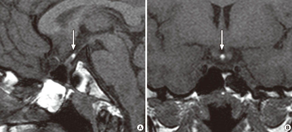
Fig. 2 Sagittal (A) and coronal (B) T1 weighted images of sella MRI show a small high signal change suggesting ectopic posterior pituitary gland (arrows). No visible normal pituitary stalk was noted.
Reference
-
1. Argente J. Diagnosis of late puberty. Horm Res. 1999. 51:Suppl 3. 95–100.2. Hopwood NJ. Pathogenesis and management of abnormal puberty. Spec Top Endocrinol Metab. 1985. 7:175–236.3. Melo ME, Marui S, Carvalho LR, Arnhold IJ, Leite CC, Mendonca BB, Knoepfelmacher M. Hormonal, pituitary magnetic resonance, LHX4 and HESX1 evaluation in patients with hypopituitarism and ectopic posterior pituitary lobe. Clin Endocrinol (Oxf). 2007. 66:95–102.4. Yoo HJ, Choi KM, Ryu OH, Suh SI, Kim NH, Baik SH, Choi DS. Delayed puberty due to pituitary stalk dysgenesis and ectopic neurohypophysis. Korean J Intern Med. 2006. 21:68–72.5. Shin SY, Kim JY, Yoon SJ, Kim SK, Hong SB, Kim YJ, Nam MS, Kim MR, Shin SP, Kim YS. A case of delayed puberty due to hypoplasia of anterior pituitary gland with pituitary stalk agenesis and ectopic neurohypophysis. J Korean Soc Endocrinol. 1999. 14:578–586.6. van der Linden AS, van Es HW. Case 112: pituitary stalk transection syndrome with ectopic posterior pituitary gland. Radiology. 2007. 243:594–597.7. Fujisawa I. Pathogenesis of an ectopic posterior lobe in patients of short stature with growth hormone deficiency. AJNR Am J Neuroradiol. 1998. 19:193–195.8. Thomas PQ, Dattani MT, Brickman JM, McNay D, Warne G, Zacharin M, Cameron F, Hurst J, Woods K, Dunger D, Stanhope R, Forrest S, Robinson IC, Beddington RS. Heterozygous HESX1 mutations associated with isolated congenital pituitary hypoplasia and septo-optic dysplasia. Hum Mol Genet. 2001. 10:39–45.9. Klose MC, Juul A, Poulsgaard L, Kosteljanetz M, Brennum J, Feldt-Rasmussen UF. [Pituitary insufficiency following head trauma]. Ugeskr Laeger. 2007. 169:211–213.10. Fujisawa I, Kikuchi K, Nishimura K, Togashi K, Itoh K, Noma S, Minami S, Sagoh T, Hiraoka T, Momoi T. Transection of the pituitary stalk: development of an ectopic posterior lobe assessed with MR imaging. Radiology. 1987. 165:487–489.11. Kikuchi K, Fujisawa I, Momoi T, Yamanaka C, Kaji M, Nakano Y, Konishi J, Mikawa H, Sudo M. Hypothalamic-pituitary function in growth hormone-deficient patients with pituitary stalk transection. J Clin Endocrinol Metab. 1988. 67:817–823.12. Pinto G, Netchine I, Sobrier ML, Brunelle F, Souberbielle JC, Brauner R. Pituitary stalk interruption syndrome: a clinical-biological-genetic assessment of its pathogenesis. J Clin Endocrinol Metab. 1997. 82:3450–3454.13. Chen S, Leger J, Garel C, Hassan M, Czernichow P. Growth hormone deficiency with ectopic neurohypophysis: anatomical variations and relationship between the visibility of the pituitary stalk asserted by magnetic resonance imaging and anterior pituitary function. J Clin Endocrinol Metab. 1999. 84:2408–2413.14. Vance ML. Hypopituitarism. N Engl J Med. 1994. 330:1651–1662.15. Ascoli P, Cavagnini F. Hypopituitarism. Pituitary. 2006. 9:335–342.16. Zamboni G, Ziviani L, Antoniazzi F, Tato L. [Empty sella syndrome: 2 cases to show its polymorphism]. Pediatr Med Chir. 1997. 19:65–67.17. Garel C, Leger J. Contribution of magnetic resonance imaging in non-tumoral hypopituitarism in children. Horm Res. 2007. 67:194–202.
- Full Text Links
-
- Actions
-
Cited
- CITED
-
- Close
- Share
-
- Similar articles
-
- A Case of Delayed Puberty due to Hypoplasia of Anterior Pituitary Gland with Pituitary Stalk Agenesis and Ectopic Neurohypophysis
- Delayed Puberty due to Pituitary Stalk Dysgenesis and Ectopic Neurohypophysis
- A Case of Hypoplastic Ectopic Kidney in Pelvic Cavity
- Diagnosing ectopic pregnancy in the emergency setting
- A case of retroperitoneal ectopic pregnancy
