Anesth Pain Med.
2016 Jan;11(1):64-67. 10.17085/apm.2016.11.1.64.
Newly detected patent ductus arteriosus by transesophageal echocardiography in patient who underwent cardiopulmonary bypass: A case report
- Affiliations
-
- 1Department of Anesthesiology and Pain Medicine, Seoul National University Hospital, Seoul, Korea. mellyn7@gmail.com
- KMID: 2169076
- DOI: http://doi.org/10.17085/apm.2016.11.1.64
Abstract
- Transesophageal echocardiography is a useful device to evaluate the posterior structure of heart with an advantage of enabling clearer images, as compared to transthoracic echocardiography. With intraoperative transesophageal echocardiography, we can reconfirm pre-diagnosed lesions, determine the success of the operation, and in particular, diagnose new lesions that are undetected in pre-operative evaluation. In the present case, undiagnosed patent ductus arteriosus was found on intraoperative transesophageal echocardiography during cardiopulmonary bypass. Subsequently, the patent ductus arteriosus was ligated successfully. With transesophageal echocardiography, we can diagnose the structural and functional abnormality of heart unidentified in the pre-operative evaluation. Also, transesophageal echocardiography can play the role of a rescuer to solve the problems that occur during cardiopulmonary bypass.
MeSH Terms
Figure
-

Fig. 1 Intraoperative transesophageal echocardiography. A 0.5 cm patent ductus arteriosus originated from descending thoracic aorta. A indicating descending thoracic aorta and arrow indicating patent ductus arteriosus.
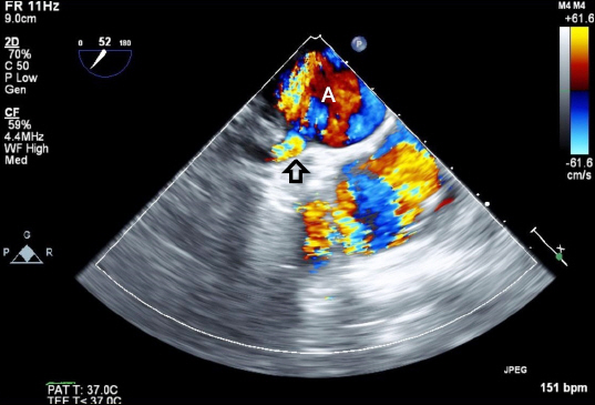
Fig. 2 Intraoperative transesophageal echocardiography with color Doppler. Blood flow in patent ductus arteriosus detected by using color Doppler. A indicating descending thoracic aorta and arrow indicating patent ductus arteriosus.
Reference
-
1. McGrath RL, McGuinness GA, Way GL, Wolfe RR, Nora JJ, Simmons MA. The silent ductus arteriosus. J Pediatr. 1978; 93:110–3. DOI: 10.1016/S0022-3476(78)80617-9.
Article2. Allen HD, Goldberg SJ, Valdes-Cruz LM, Sahn DJ. Use of echocardiography in newborns with patent ductus arteriosus: a review. Pediatr Cardiol. 1982; 3:65–70. DOI: 10.1007/BF02082335. PMID: 7155942.
Article3. Houston AB, Gnanapragasam JP, Lim MK, Doig WB, Coleman EN. Doppler ultrasound and the silent ductus arteriosus. Heart. 1991; 65:97–9. DOI: 10.1136/hrt.65.2.97.
Article4. Marelli AJ, Child JS, Perloff JK. Transesophageal echocardiography in congenital heart disease in the adult. Cardiol Clin. 1993; 11:505–20. PMID: 8402777.
Article5. Chugh R, Salem MM. Echocardiography for patent ductus arteriosus including closure in adults. Echocardiography. 2015; 32(Suppl 2):S125–39. DOI: 10.1111/echo.12457. PMID: 24888537.
Article6. Shyu KG, Lai LP, Lin SC, Chang H, Chen JJ. Diagnostic accuracy of transesophageal echocardiography for detecting patent ductus arteriosus in adolescents and adults. Chest. 1995; 108:1201–5. DOI: 10.1378/chest.108.5.1201. PMID: 7587417.
Article7. Cheitlin MD, Armstrong WF, Aurigemma GP, Beller GA, Bierman FZ, Davis JL, et al. ACC/AHA/ASE 2003 guideline update for the clinical application of echocardiography. A Report of the American College of Cardiology/American Heart Association Task Force on Practice Guidelines (ACC/AHA/ASE Committee to Update the 1997 Guidelines for the Clinical Application of Echocardiography). J Am Coll Cardiol. 2003; 42:954–70. DOI: 10.1016/S0735-1097(03)01065-9.8. American Society of Anesthesiologists and Society of Cardiovascular Anesthesiologists Task Force on Transesophageal Echocardiography. Practice guidelines for perioperative transesophageal echocardiography. An updated report by the American Society of Anesthesiologists and the Society of Cardiovascular Anesthesiologists Task Force on Transesophageal Echocardiography. Anesthesiology. 2010; 112:1084–96. PMID: 20418689.9. Gogia R, Kumar B, Jayant A. A proposed method to visualize the ductus arteriosus on transesophageal echocardiography. Ann Card Anaesth. 2014; 17:296–8. DOI: 10.4103/0971-9784.142068. PMID: 25281628.
Article10. Kanter KR, Schaff HV, Gott VL, Gardner TJ. Reduced oxygen consumption with effective left ventricular venting during postischemic reperfusion. Circulation. 1982; 66:I50–4. PMID: 7083546.
- Full Text Links
-
- Actions
-
Cited
- CITED
-
- Close
- Share
-
- Similar articles
-
- Surgical Treatment of Persistent Ductus Arteriosus Complicated by Bacterial Endocarditis with Pulmonary Artery Embolism: 1 case report
- Nonsurgical closure of patent ductus arteriosus with the rashkind PDA occluder system
- Three Cases of Hemolysis After Transcatheter Closure of A Patent Ductus Arteriosus
- Aneurysm after Surgical Ligation of Patent Ductus Arteriosus: A Case Report
- A case of congenital ductus arteriosus aneurysm
