Cancer Res Treat.
2011 Jun;43(2):131-133.
Benign Metastasizing Leiomyoma with Multiple Lymph Node Metastasis: A Case Report
- Affiliations
-
- 1Department of Obstetrics and Gynecology, Samsung Medical Center, Sungkyunkwan University School of Medicine, Seoul, Korea. bgkim@skku.edu
- 2Department of Pathology, Samsung Medical Center, Sungkyunkwan University School of Medicine, Seoul, Korea.
Abstract
- This is a case report about benign metastasizing leiomyoma with multiple lymph node metastasis. A 34-year-old woman received an abdominal myomectomy for a suspicious leiomyoma. On the pathology report, atypical leiomyoma was suspected. Due to the suspicion of multiple lymph node metastasis on pelvis computed tomography (CT) 1 year after the operation, she was transferred to the Samsung Medical Center on October, 2009 for further work up. According to original slide review, it was determined to be a benign leiomyoma with a mitotic count <5/10 high-power fields, little cytological atypia and no tumor cell necrosis. Additional immunostaining was done. Multiple lymph node metastasis and a small lung nodule were identified on positron emission tomogarphy-CT and chest CT. Extensive debulking surgery and diagnostic video-assisted thoracoscopic surgery (VATS) wedge resection were subsequently done. Metastatic lesions were reported to have a histology similar to that of the original mass. VATS right upper lobectomy with mediastinal lymph node dissection was performed because of the pathology result of VATS (adenocarcinoma). She started taking an aromatase inhibitor (Letrozole(R)) and there was no evidence of recurrence of disease on an imaging study and no post-operative complications until recently.
MeSH Terms
Figure
-

Fig. 1 Original mass from a myomectomy specimen composed of benign looking spindle cells (H&E, ×200).
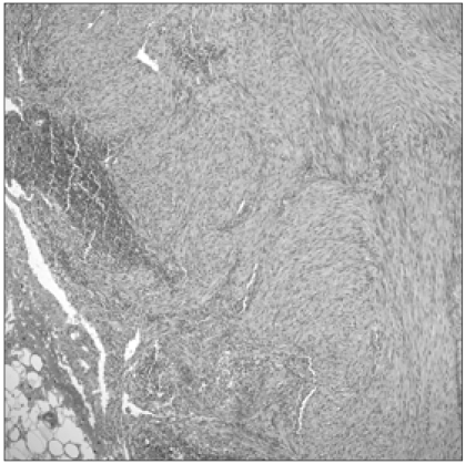
Fig. 2 Lymph node metastasis (para-aortic lymph node) (H&E, ×100).
Reference
-
1. Hendrickson MR, Kempson RL. Surgical pathology of the uterine corpus. Major Probl Pathol. 1979; 12:1–580. PMID: 537399.2. Tietze L, Günther K, Hörbe A, Pawlik C, Klosterhalfen B, Handt S, et al. Benign metastasizing leiomyoma: a cytogenetically balanced but clonal disease. Hum Pathol. 2000; 31:126–128. PMID: 10665925.
Article3. Egberts JH, Schafmayer C, Bauerschlag DO, Jänig U, Tepel J. Benign abdominal and pulmonary metastasizing leiomyoma of the uterus. Arch Gynecol Obstet. 2006; 274:319–322. PMID: 16649038.
Article4. Kayser K, Zink S, Schneider T, Dienemann H, André S, Kaltner H, et al. Benign metastasizing leiomyoma of the uterus: documentation of clinical, immunohistochemical and lectin-histochemical data of ten cases. Virchows Arch. 2000; 437:284–292. PMID: 11037349.
Article
- Full Text Links
-
- Actions
-
Cited
- CITED
-
- Close
- Share
-
- Similar articles
-
- A Case of Benign Metastasizing Leiomyoma of Uterus to the Lung
- A Case of Metastasizing Pleomorphic Adenoma Recurred as Cervical Lymph Node Metastasis after Parotidectomy
- A Case of Metastasis to Lung of Benign Metastasizing Leiomyoma
- Unusual CT Findings of a Benign Metastasizing Leiomyoma Presenting with Multiple Cavitary Nodules: A Case Report
- Multiple benign metastasizing leiomyomas in the pelvic and para-aortic lymph nodes: A case report
