CBCT findings of periapical cemento-osseous dysplasia: A case report
- Affiliations
-
- 1Department of Oral and Maxillofacial Radiology, Faculty of Dentistry, Hamadan University of Medical Science, Hamadan, Iran. faezehyousefi@yahoo.com
- KMID: 2167461
- DOI: http://doi.org/10.5624/isd.2013.43.3.215
Abstract
- Periapical cemento-osseous dysplasia (PCOD) is a subtype of cemento-osseous dysplasia that usually occurs in middle-aged black women. This report described a case of a 45-year-old Iranian woman who was diagnosed with PCOD on the basis of cone beam computed tomographic (CBCT) findings. CBCT enabled detailed visualization of the bone changes. This report described the special radiographic characteristics of PCOD, including discontinuity of the lingual cortex on the CBCT sectional and three-dimensional images.
MeSH Terms
Figure
-
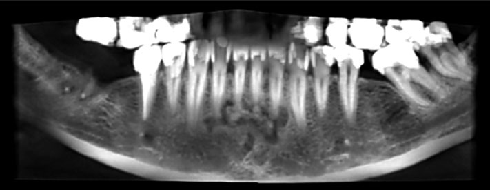
Fig. 1 A reformatted panoramic CBCT image shows a multifocal lesion extending from the mesial side of the right mandibular lateral incisor to the distal side of the left mandibular lateral incisor.
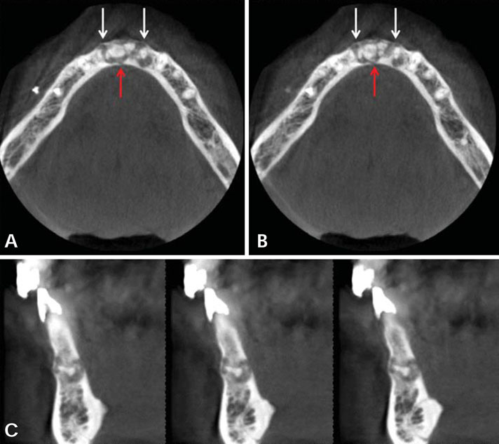
Fig. 2 A. An axial CBCT image shows the expansion of the buccal cortex (white arrows) and discontinuity of the lingual cortex (red arrow). B. Another axial CBCT image shows the same features as A. C. Cross-sectional CBCT images show the discontinuity of the lingual cortical plate.
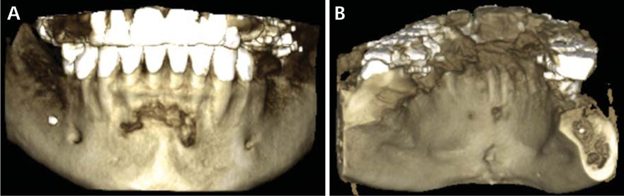
Fig. 3 Three-dimensional reconstructed CBCT images of the mandible. A. The frontal view shows erosion of the buccal cortex of the lesion. B. The lingual view shows that the lesion erodes the lingual cortex.

Fig. 4 A periapical radiograph shows a mixed radiolucent-radiopaque appearance of the lesion, located on the apices of the lower incisors. The lamina dura surrounding the apical areas of the involved teeth is lost.
Cited by 2 articles
-
Radiolucent rim as a possible diagnostic aid for differentiating jaw lesions
Hamed Mortazavi, Maryam Baharvand, Somayeh Rahmani, Soudeh Jafari, Parvin Parvaei
Imaging Sci Dent. 2015;45(4):253-261. doi: 10.5624/isd.2015.45.4.253.Recurrent symptomatic cemento-osseous dysplasia: A case report
Chang-Ki Min, Kwang-Joon Koh, Kyoung-A Kim
Imaging Sci Dent. 2018;48(2):131-137. doi: 10.5624/isd.2018.48.2.131.
Reference
-
1. Eversole R, Su L, ElMofty S. Benign fibro-osseous lesions of the craniofacial complex. A review. Head Neck Pathol. 2008; 2:177–202.
Article2. Thakkar N, Horner K, Sloan P. Familial occurrence of periapical cemental dysplasia. Virchows Arch A Pathol Anat Histopathol. 1993; 423:233–236.
Article3. Kawai T, Hiranuma H, Kishino M, Jikko A, Sakuda M. Cemento-osseous dysplasia of the jaws in 54 Japanese patients: a radiographic study. Oral Surg Oral Med Oral Pathol Oral Radiol Endod. 1999; 87:107–114.4. Komabayashi T, Zhu Q. Cemento-osseous dysplasia in an elderly Asian male: a case report. J Oral Sci. 2011; 53:117–120.
Article5. Falace D, Cunningham C. Periapical cemental dysplasia: simultaneous occurrence in multiple maxillary and mandibular teeth. J Endod. 1984; 10:455–456.
Article6. Alsufyani NA, Lam EW. Cemento-osseous dysplasia of the jaw bones: key radiographic features. Dentomaxillofac Radiol. 2011; 40:141–146.
Article7. DiFiore P, Bowen S. Cemento-osseous dysplasia in African-American men: a report of two clinical cases. J Tenn Dent Assoc. 2010; 90:26–29.8. MacDonald-Jankowski DS. Florid cemento-osseous dysplasia: a systematic review. Dentomaxillofac Radiol. 2003; 32:141–149.
Article9. Zegarelli E, Kutscher A, Napoli N, Iurono F, Hoffman P. The cementoma. A study of 230 patients with 435 cementomas. Oral Surg Oral Med Oral Pathol. 1964; 17:219–224.10. Scholl RJ, Kellett HM, Neumann DP, Lurie AG. Cysts and cystic lesions of the mandible: clinical and radiologic-histopathologic review. Radiographics. 1999; 19:1107–1124.
Article11. Manganaro AM, Millett GV. Periapical cemental dysplasia. Gen Dent. 1996; 44:336–339.12. Alawi F. Benign fibro-osseous diseases of the maxillofacial bones. A review and differential diagnosis. Am J Clin Pathol. 2002; 118:Suppl. S50–S70.13. Alsufyani NA, Lam EW. Osseous (cemento-osseous) dysplasia of the jaws: clinical and radiographic analysis. J Can Dent Assoc. 2011; 77:b70.14. Macdonald-Jankowski DS. Focal cemento-osseous dysplasia: a systematic review. Dentomaxillofac Radiol. 2008; 37:350–360.
Article15. Ariji Y, Ariji E, Higuchi Y, Kubo S, Nakayama E, Kanda S. Florid cemento-osseous dysplasia. Radiographic study with special emphasis on computed tomography. Oral Surg Oral Med Oral Pathol. 1994; 78:391–396.
- Full Text Links
-
- Actions
-
Cited
- CITED
-
- Close
- Share
-
- Similar articles
-
- 3 Types of Cemento-Osseous Dysplasia: Case Reports
- Florid cemento-osseous dysplasia: a report of two cases
- Misdiagnosis of florid cemento-osseous dysplasia leading to unnecessary root canal treatment: a case report
- Recurrent symptomatic cemento-osseous dysplasia: A case report
- Cemento-Osseous Dysplasia Largely Occurring in the Mandible: Case Report
