Attitude of the Korean dentists towards radiation safety and selection criteria
- Affiliations
-
- 1Department of Oral and Maxillofacial Radiology and Wonkwang Dental Research Institute, College of Dentistry, Wonkwang University, Iksan, Korea.
- 2Graduate Program in Oral and Maxillofacial Radiology, School of Dentistry, University of North Carolina, Chapel Hill, NC, USA. ludlowj@dentistry.unc.edu
- KMID: 2167455
- DOI: http://doi.org/10.5624/isd.2013.43.3.179
Abstract
- PURPOSE
X-ray exposure should be clinically justified and each exposure should be expected to give patients benefits. Since dental radiographic examination is one of the most frequent radiological procedures, radiation hazard becomes an important public health concern. The purpose of this study was to investigate the attitude of Korean dentists about radiation safety and use of criteria for selecting the frequency and type of radiographic examinations.
MATERIALS AND METHODS
The study included 267 Korean dentists. Five questions related to radiation safety were asked of each of them. These questions were about factors associated with radiation protection of patients and operators including the use of radiographic selection criteria for intraoral radiographic procedures.
RESULTS
The frequency of prescription of routine radiographic examination (an example is a panoramic radiograph for screening process for occult disease) was 34.1%, while that of selective radiography was 64.0%. Dentists' discussion of radiation risk and benefit with patients was infrequent. More than half of the operators held the image receptor by themselves during intraoral radiographic examinations. Lead apron/thyroid collars for patient protection were used by fewer than 22% of dental offices. Rectangular collimation was utilized by fewer than 15% of dental offices.
CONCLUSION
The majority of Korean dentists in the study did not practice radiation protection procedures which would be required to minimize exposure to unnecessary radiation for patients and dental professionals. Mandatory continuing professional education in radiation safety and development of Korean radiographic selection criteria is recommended.
Keyword
MeSH Terms
Figure
-
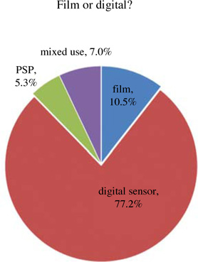
Fig. 1 Frequency of patient's questions about the radiation hazard.
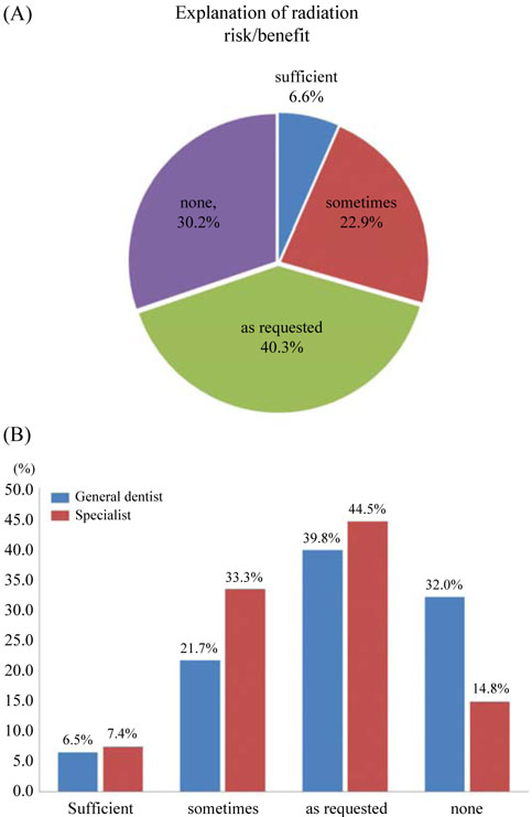
Fig. 2 A. Frequency of dentist explanation about radiation risk/benefit to patients. B. Comparison of frequency of explaining about radiation risk/benefit to patients, between general dentists and specialists of oral radiologist. "Sometimes": depending on situation, "as requested": when patients ask.
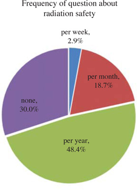
Fig. 3 Frequency of image receptor usage among the film, digital sensor, photostimulable phosphor (PSP) plate. Mixed means "film and digital combination".
Cited by 1 articles
-
Insights into the state of radiation protection among a subpopulation of Indian dental practitioners
Almas Binnal, Gururaghavendran Rajesh, Ceena Denny, Junaid Ahmed, Vijayendra Nayak
Imaging Sci Dent. 2013;43(4):253-259. doi: 10.5624/isd.2013.43.4.253.
Reference
-
1. Preston-Martin S, White SC. Brain and salivary gland tumors related to prior dental radiography: implications for current practice. J Am Dent Assoc. 1990; 120:151–158.
Article2. Memon A, Godward S, Williams D, Siddique I, Al-Saleh K. Dental x-rays and the risk of thyroid cancer: a case-control study. Acta Oncol. 2010; 49:447–453.
Article3. Claus EB, Calvocoressi L, Bondy ML, Schildkraut JM, Wiemels JL, Wrensch M. Dental x-rays and risk of meningioma. Cancer. 2012; 118:4530–4537.
Article4. Martinez Beneyto Y, Alcaraz Banos M, Perez Lajarin L, Rushton VE. Clinical justification of dental radiology in adult patients: a review of the literature. Med Oral Patol Oral Cir Bucal. 2007; 12:E244–E251.5. Frederiksen NL. Guidelines for prescribing dental radiographs. United States Food and Drug Administration. Tex Dent J. 1995; 112:63–67.6. Stephens RG, Kogon SL. New U.S. guidelines for prescribing dental radiographs - a critical review. J Can Dent Assoc. 1990; 56:1019–1024.7. Kim IH, Mupparapu M. Dental radiographic guidelines: a review. Quintessence Int. 2009; 40:389–398.8. Horner K, Islam M, Flygare L, Tsiklakis K, Whaites E. Basic principles for use of dental cone beam computed tomography: consensus guidelines of the European Academy of Dental and Maxillofacial Radiology. Dentomaxillofac Radiol. 2009; 38:187–195.
Article9. Tyndall DA, Price JB, Tetradis S, Ganz SD, Hildebolt C, Scarfe WC. Position statement of the American Academy of Oral and Maxillofacial Radiology on selection criteria for the use of radiology in dental implantology with emphasis on cone beam computed tomography. Oral Surg Oral Med Oral Pathol Oral Radiol. 2012; 113:817–826.
Article10. Ilgüy D, Ilgüy M, Dinçer S, Bayirli G. Survey of dental radiological practice in Turkey. Dentomaxillofac Radiol. 2005; 34:222–227.11. American Academy of Pediatric Dentistry, Ad Hoc Committee on Pedodontic Radiology. Guideline on prescribing dental radiographs for infants, children, adolescents, and persons with special health care needs. Pediatr Dent. 2012; 34:189–191.12. Kantor ML. Longitudinal trends in the use of individualized radiographic examinations at dental schools in the United States and Canada. J Dent Educ. 2006; 70:160–168.
Article13. American Dental Association [Internet]. The selection of patients for dental radiographic examinations. Revised 2004. cited 2013 Jan 15. Available from https://www.ada.org/sections/advocacy/pdfs/topics_radiography_examinations(2).pdf.14. Matteson SR, Joseph LP, Bottomley W, Finger HW, Frommer HH, Koch RW, et al. The report of the panel to develop radiographic selection criteria for dental patients. Gen Dent. 1991; 39:264–270.15. ICRP. Recommendations of the ICRP. ICRP Publication 26. Ann ICRP. 1977; 1:1–53.16. American Dental Association Council on Scientific Affairs. The use of dental radiographs: update and recommendations. J Am Dent Assoc. 2006; 137:1304–1312.17. Kantor ML. Use of radiology practice guidelines and compliance with accreditation standards in US and Canadian dental schools. J Dent Res. 2000; 79:1532–1536.
Article18. Atchison KA, White SC, Flack VF, Hewlett ER, Kinder SA. Efficacy of the FDA selection criteria for radiographic assessment of the periodontium. J Dent Res. 1995; 74:1424–1432.
Article19. Atchison KA, White SC, Flack VF, Hewlett ER. Assessing the FDA guidelines for ordering dental radiographs. J Am Dent Assoc. 1995; 126:1372–1383.
Article20. Recommendations in radiographic practices, 1984. Council on Dental Materials, Instruments, and Equipment. J Am Dent Assoc. 1984; 109:764–765.21. McCarley DH. ADA Principles of Ethics and Code of Professional Conduct. Tex Dent J. 2011; 128:728–732.22. Goodman JM. Protect yourself! Make a plan to obtain "informed refusal". OBG Management. 2007; 19:45–50.23. American Association of Endodontists. American Acadamey of Oral and Maxillofacial Radiography. AAE and AAOMR joint position statement. Use of cone-beam-computed tomography in endodontics. Pa Dent J (Harrisb). 2011; 78:37–39.24. de Araujo FB, de Araujo DR, dos Santos CK, de Souza MA. Diagnosis of approximal caries in primary teeth: radiographic versus clinical examination using tooth separation. Am J Dent. 1996; 9:54–56.25. Pitts NB. The use of bitewing radiographs in the management of dental caries: scientific and practical considerations. Dentomaxillofac Radiol. 1996; 25:5–16.
Article26. Gibbs SJ, Pujol A Jr, Chen TS, Carlton JC, Dosmann MA, Malcolm AW, et al. Radiation doses to sensitive organs from intraoral dental radiography. Dentomaxillofac Radiol. 1987; 16:67–77.
Article27. NCRP. Report No. 145. Radiation protection in dentistry. Bethesda: National Council on Radiation Protection and Measurements;2003.28. Hendry JH, Simon SL, Wojcik A, Sohrabi M, Burkart W, Cardis E, et al. Human exposure to high natural background radiation: what can it teach us about radiation risks? J Radiol Prot. 2009; 29:A29–A42.
Article29. Hujoel PP, Bollen AM, Noonan CJ, del Aguila MA. Antepartum dental radiography and infant low birth weight. JAMA. 2004; 291:1987–1993.
Article30. Sikorski PA, Taylor KW. The effectiveness of the thyroid shield in dental radiology. Oral Surg Oral Med Oral Pathol. 1984; 58:225–236.
Article31. Araki K, Kanda S. Radiological characteristics of lead foils in dental film packets: analysis of components and shielding effect. Dentomaxillofac Radiol. 1992; 21:21–25.
Article32. White SC, Pharoah MJ. Oral radiology: principles and interpretation. 6th ed. St Louis: Mosby;2009.33. Parrott LA, Ng SY. A comparison between bitewing radiographs taken with rectangular and circular collimators in UK military dental practices: a retrospective study. Dentomaxillofac Radiol. 2011; 40:102–109.
Article34. Svenson B, Söderfeldt B, Gröndahl HG. Attitudes of Swedish dentists to the choice of dental X-ray film and collimator for oral radiology. Dentomaxillofac Radiol. 1996; 25:157–161.
Article35. Jacobs R, Vanderstappen M, Bogaerts R, Gijbels F. Attitude of the Belgian dentist population towards radiation protection. Dentomaxillofac Radiol. 2004; 33:334–339.
Article36. Bohay RN, Kogon SL, Stephens RG. A survey of radiographic techniques and equipment used by a sample of general dental practitioners. Oral Surg Oral Med Oral Pathol. 1994; 78:806–810.
Article37. Geist JR, Katz JO. The use of radiation dose-reduction techniques in the practices of dental faculty members. J Dent Educ. 2002; 66:697–702.
Article38. Orafi I, Rushton VE. A questionnaire study to derive information on the working environment, clinical training, use of ancillary staff and optimization of patient radiation dose within UK dental practice. Int Endod J. 2012; 45:763–772.
Article
- Full Text Links
-
- Actions
-
Cited
- CITED
-
- Close
- Share
-
- Similar articles
-
- Status of Mongolian dentistry viewed from information resources and selection of adhesive dental restorative materials and continuing education
- Radiation protection in dental clinic
- Factors Influencing Radiation Protection Behaviors of Endoscopy Nurses during Endoscopic Interventional Radiology
- Comparison of Musculoskeletal Disorders between Pediatric Dentists and General Dentists
- Dentists’ Recognition of Child Abuse and Neglect and Mandatory Attitude to Report
