The arterial blood supply of the temporomandibular joint: an anatomical study and clinical implications
- Affiliations
-
- 1Department of Surgical and Oncological Disciplines, University of Palermo, Palermo, Italy. cucciaam@odonto.unipa.it
- 2Department of Biomorphology and Biotechnologies, University of Messina, Messina, Italy.
- 3Villa Santa Teresa, Diagnostica per Immagini, Palermo, Italy.
- KMID: 2167445
- DOI: http://doi.org/10.5624/isd.2013.43.1.37
Abstract
- PURPOSE
The aim of this study was to analyze three-dimensional images of the arterial supply to the temporomandibular joint.
MATERIALS AND METHODS
Ten patients (five men and five women, mean age 36 years) without signs or symptoms of temporomandibular disorders, who underwent contrast-enhanced computed tomographic (CT) scanning with intravenous contrast, were studied. The direct volume rendering technique of CT images was used, and a data set of images to visualize the vasculature of the human temporomandibular joint in three dimensions was created. After elaboration of the data through post-processing, the arterial supply of the temporomandibular joint was studied.
RESULTS
The analysis revealed the superficial temporal artery, the anterior tympanic artery, the deep temporal artery, the auricular posterior artery, the transverse facial artery, the middle meningeal artery, and the maxillary artery with their branches as the main arterial sources for the lateral and medial temporomandibular joint.
CONCLUSION
The direct volume rendering technique was found to be successful in the assessment of the arterial supply to the temporomandibular joint. The superficial temporal artery and maxillary artery ran along the lateral and medial sides of the condylar neck, suggesting that these arteries are at increased risk during soft-tissue procedures such as an elective arthroplasty of the temporomandibular joint.
Keyword
MeSH Terms
Figure
-
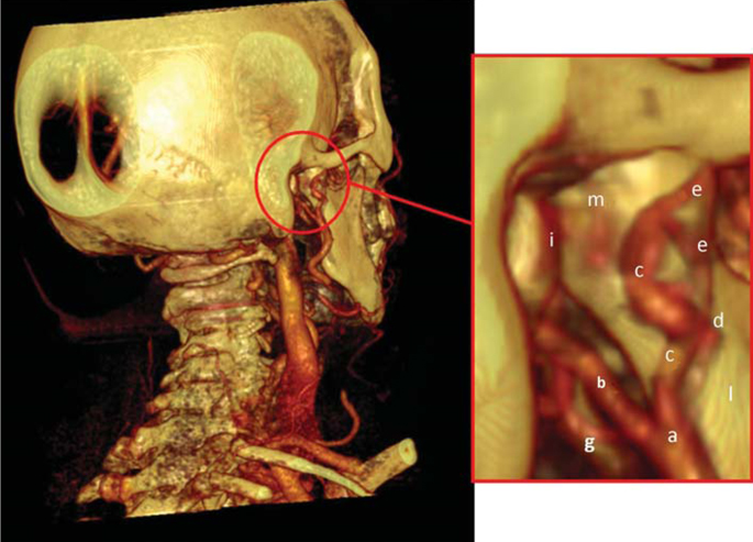
Fig. 1 Posterior view of computerized tomography rendering of the temporomandibular joint of a 30-year-old healthy female. The external carotid artery (a), the internal maxillary artery (b), the superficial temporal artery (c), the transverse facial artery (d), with their small diverging arteries (e), the middle meningeal artery (g), the retrodiscal tissue (i), the ramus (l) and the condyle (m)

Fig. 2 Posterior and lateral view of computerized tomography rendering of the temporomandibular joint of a 30-year-old healthy female. The external carotid artery (a), the internal maxillary artery (b), the superficial temporal artery (c), the transverse facial artery (d), the inferior dental artery (f), the middle meningeal artery (g), the anterior tympanic artery (h), the retrodiscal tissue (i), the ramus (l) and the condyle (m), the temporal posterior artery (t)
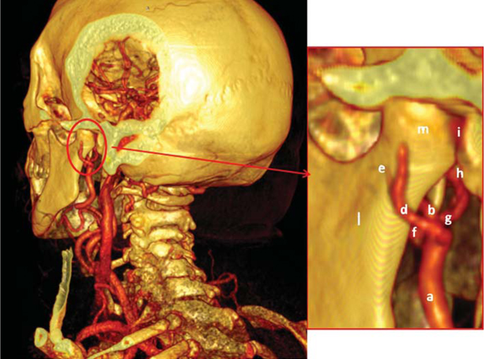
Fig. 3 Posterior view of computerized tomography rendering of the temporomandibular joint of a 30-year-old healthy female. The external carotid artery (a), the internal maxillary artery (b), the transverse facial artery (d) with their small diverging arteries (e), the inferior dental artery (f), the middle meningeal artery (g), the anterior timpanic artery (h), the retrodiscal tissue (i), the ramus (l) and the condyle (m)

Fig. 4 Computerized tomography rendering of the left temporomandibular joint of a 28-year-old healthy male (A) and diagram (B) show the branches of maxillary artery. The external carotid artery (a), the internal maxillary artery (b), the inferior dental artery (f), the middle meningeal artery (g), the condyle (m), the masseteric artery (n), the pterygoid artery (o), the sphenopalatine artery (p), the sphenopalatine foramen (q), the deep temporal artery (v)

Fig. 5 Lateral angiograms (A) and diagrams (B and C) of the internal maxillary artery and the superficial temporal artery arising from the external carotid artery. The external carotid artery (a), the internal maxillary artery (b), the superficial temporal artery (c), the transverse facial artery (d), the inferior dental artery (f), the masseteric artery (n), the pterygoid artery (o), the sphenopalatine artery (p), the occipital artery (r), the auricular posterior artery (s), the temporal posterior artery (t), the deep temporal anterior artery (u), the deep temporal artery (v)
Reference
-
1. Molinari F, Manicone PF, Raffaelli L, Raffaelli R, Pirronti T, Bonomo L. Temporomandibular joint soft-tissue pathology, I: Disc abnormalities. Semin Ultrasound CT MR. 2007. 28:192–204.
Article2. Patnaik VV, Bala S, Singla RK. Anatomy of temporomandibular joint? A review. J Anat Soc India. 2000. 49:191–197.3. Ezure H, Mori R, Ito J, Otsuka N. Case of a completely absent facial artery. Int J Anat Var. 2011. 4:72–74.4. Putz R, Pabst R. Sobotta atlas of human anatomy. Vol. 1: head, neck, upper limb. 2001. 13 rev. ed. Munich: Urban & Fischer;86–87.5. Hatcher DC, Blom RJ, Baker CG. Temporomandibular joint spatial relationships: osseous and soft tissues. J Prosthet Dent. 1986. 56:344–353.
Article6. Takagi R, Shimoda T, Westesson PL, Takahashi A, Morris TW, Sano T, et al. Angiography of the temporomandibular joint. Description of an experimental technique with initial results. Oral Surg Oral Med Oral Pathol. 1994. 78:539–543.7. Piette E, Lametschwandtner A. The angioarchitecture of the rat mandibular joint synovium. Arch Oral Biol. 1995. 40:487–497.
Article8. Piette E, Lametschwandtner A. The angioarchitecture of the rat mandibular joint bilaminar zone. Arch Oral Biol. 1995. 40:499–505.
Article9. Piette E, Lametschwandtner A. The fine vasculature of the rat mandibular joint. Acta Anat (Basel). 1995. 153:64–72.
Article10. Kvinnsland S, Kvinnsland I, Kristiansen AB. Effect of experimental traumatic occlusion on blood flow in the temporomandibular joint of the rat. Acta Odontol Scand. 1993. 51:293–298.
Article11. Mérida Velasco JR, Rodríguez Vázquez JF, Jiménez Collado J. Anterior tympanic artery: course, ramification and relationship with the temporomandibular joint. Acta Anat (Basel). 1997. 158:222–226.
Article12. Pehling J, Schiffman E, Look J, Shaefer J, Lenton P, Fricton J. Interexaminer reliability and clinical validity of the temporomandibular index: a new outcome measure for temporomandibular disorders. J Orofac Pain. 2002. 16:296–304.13. Huskisson EC. Measurement of pain. Lancet. 1974. 2:1127–1131.
Article14. Takagi R, Westesson PL, Ohashi Y, Togashi H. MR angiography of the TMJ in asymptomatic volunteers. Oral Radiol. 1998. 14:69–74.
Article15. Uysal II, Buyukmumcu M, Dogan NU, Seker M, Ziylan T. Clinical significance of maxillary artery and its branches: a cadaver study and review of the literature. Int J Morphol. 2011. 29:1274–1281.
Article16. Boyer CC, Williams W, Stevens FH. Blood supply of the temporomandibular joint. J Dent Res. 1964. 43:224–228.
Article17. Funakoshi K. Nutrient arteries of the temporomandibular joint: an anatomical and a pathological study. Okajimas Folia Anat Jpn. 2001. 78:7–16.18. Sparacia G, Bencivinni F, Banco A, Sarno C, Bartolotta TV, Lagalla R. Imaging processing for CT angiography of the cervicocranial arteries: evaluation of reformatting technique. Radiol Med. 2007. 112:224–238.
Article19. Benvenuti L, Chibbaro S, Carnesecchi S, Pulera F, Gagliardi R. Automated three-dimensional volume rendering of helical computed tomographic angiography for aneurysms: an advanced application of neuronavigation technology. Neurosurgery. 2005. 57:1 Suppl. 69–77.
Article20. Ferretti GR, Arbib F, Bertrand B, Coulomb M. Haemoptysis associated with pulmonary varices: demonstration using computed tomographic angiography. Eur Respir J. 1998. 12:989–992.
Article21. Godlewski G, Bossy J, Giraudon M, Dussaud J, Pavart JC, Lopez JF. Arterial vascularization of the temporomandibular joint. Bull Assoc Anat (Nancy). 1978. 62:229–236.22. Siéssere S, Vitti M, Semprini M, Regalo SC, Iyomasa MM, Dias FJ, et al. Macroscopic and microscopic aspects of the temporomandibular joint related to its clinical implication. Micron. 2008. 39:852–858.
Article23. Wasicky R, Pretterklieber ML. The human anterior tympanic artery. A nutrient artery of the middle ear with highly variable origin. Cells Tissues Organs. 2000. 166:388–394.24. Cillo JE, Sinn D, Truelson JM. Management of middle meningeal and superficial temporal artery hemorrhage from total temporomandibular joint replacement surgery with a gelatinbased hemostatic agent. J Craniofac Surg. 2005. 16:309–312.
Article25. Rajab BM, Sarraf AA, Abubaker AO, Laskin DM. Masseteric artery: anatomic location and relationship to the temporomandibular joint area. J Oral Maxillofac Surg. 2009. 67:369–371.
Article26. Holmlund A, Hellsing G. Arthroscopy of the TMJ. An autopsy study. Int J Oral Surg. 1985. 14:169–175.27. Talebzadeh N, Rosenstein TP, Pogrel MA. Anatomy of the structures medial to the temporomandibular joint. Oral Surg Oral Med Oral Pathol Oral Radiol Endod. 1999. 88:674–678.
Article28. Weinberg LA. The etiology, diagnosis, and treatment of TMJ dysfunction-pain syndrome. Part II: Differential diagnosis. J Prosthet Dent. 1980. 43:58–70.
Article29. Tallents RH, Macher DJ, Kyrkanides S, Katzberg RW, Moss ME. Prevalence of missing posterior teeth and intraarticular temporomandibular disorders. J Prosthet Dent. 2002. 87:45–50.
Article30. Heffez LB, Jordan SL. Superficial vascularity of temporomandibular joint retrodiskal tissue: an element of the internal derangement process. Cranio. 1992. 10:180–191.
Article31. Goffinet L, Laure B, Tayeb T, Amado D, Herbreteau D, Arbeille P, et al. An arteriovenous fistula of the maxillary artery as a complication of Le Fort I osteotomy. J Craniomaxillofac Surg. 2010. 38:251–254.
Article32. Manning MP, Marshall JH. Aneurysm after arthroscopy. J Bone Joint Surg Br. 1987. 69:151.
Article33. Stewart CL, Cohen-Kerem R, Ngan BY, Forte V. Post-traumatic facial artery aneurysm in a child. Int J Pediatr Otorhinolaryngol. 2004. 68:1539–1543.
Article34. Conner WC 3rd, Rohrich RJ, Pollock RA. Traumatic aneurysms of the face and temple: a patient report and literature review, 1644 to 1998. Ann Plast Surg. 1998. 41:321–326.35. Bozkurt M, Kapi E, Karakol P, Yorgancilar E. Sudden rupture of the internal maxillary artery causing pseudoaneurysm (mandibular part) secondary to subcondylar mandible fracture. J Craniofac Surg. 2009. 20:1430–1432.
Article36. Walker MT, Liu BP, Salehi SA, Badve S, Batjer HH. Superficial temporal artery pseudoaneurysm: diagnosis and preoperative planning with CT angiography. AJNR Am J Neuroradiol. 2003. 24:147–150.37. Suzuki S, Kimura Y, Kanaji M, Sudo M, Igarashi M, Yamamoto H. Traumatic maxillary artery aneurysm with rupture into the maxillary sinus. Pract Otol (Kyoto). 1999. 92:1107–1110.
Article38. Mauldin FW, Cornay WJ 3rd, Mahaley MS Jr, Hicks JN. Severe epistaxis from a false aneurysm of the external carotid artery. Otolaryngol Head Neck Surg. 1989. 101:588–590.
Article
- Full Text Links
-
- Actions
-
Cited
- CITED
-
- Close
- Share
-
- Similar articles
-
- Investigation of the effects of temporomandibular joint arthrocentesis on blood volume of the retinal structures
- A study on simultation of the mandibular movement of the patients with temporomandibular joint disorder
- Autogenous auricular cartilage graft for repair of temporomandibular joint disk
- Unrecognized Bilateral Dislocation of Temporomandibular Joint during Orotracheal Intubation
- A study on the occlusal contact of the subjects with temporomandibular joint sound
