Development of 3D statistical mandible models for cephalometric measurements
- Affiliations
-
- 1Department of Oral and Maxillofacial Radiology, School of Dentistry, Seoul National University, Seoul, Korea.
- 2Department of Oral and Maxillofacial Radiology, BK21, and Dental Research Institute, School of Dentistry, Seoul National University, Seoul, Korea. wjyi@snu.ac.kr
- 3Department of Oral and Maxillofacial Surgery, BK21, and Dental Research Institute, School of Dentistry, Seoul National University, Seoul, Korea.
- 4Department of Periodontology, BK21, and Dental Research Institute, School of Dentistry, Seoul National University, Seoul, Korea.
- 5Division of Multimedia Engineering, Seoul Women's University, Seoul, Korea.
- KMID: 2167436
- DOI: http://doi.org/10.5624/isd.2012.42.3.175
Abstract
- PURPOSE
The aim of this study was to provide sex-matched three-dimensional (3D) statistical shape models of the mandible, which would provide cephalometric parameters for 3D treatment planning and cephalometric measurements in orthognathic surgery.
MATERIALS AND METHODS
The subjects used to create the 3D shape models of the mandible included 23 males and 23 females. The mandibles were segmented semi-automatically from 3D facial CT images. Each individual mandible shape was reconstructed as a 3D surface model, which was parameterized to establish correspondence between different individual surfaces. The principal component analysis (PCA) applied to all mandible shapes produced a mean model and characteristic models of variation. The cephalometric parameters were measured directly from the mean models to evaluate the 3D shape models. The means of the measured parameters were compared with those from other conventional studies. The male and female 3D statistical mean models were developed from 23 individual mandibles, respectively.
RESULTS
The male and female characteristic shapes of variation produced by PCA showed a large variability included in the individual mandibles. The cephalometric measurements from the developed models were very close to those from some conventional studies.
CONCLUSION
We described the construction of 3D mandibular shape models and presented the application of the 3D mandibular template in cephalometric measurements. Optimal reference models determined from variations produced by PCA could be used for craniofacial patients with various types of skeletal shape.
MeSH Terms
Figure
-

Fig. 1 Decomposition of 3D mandible shape for the construction of correspondence maps.

Fig. 2 Established landmarks for 3D cephalometric measurements in different viewing positions.

Fig. 3 Male and female 3D statistical mean models (left: male, right: female).
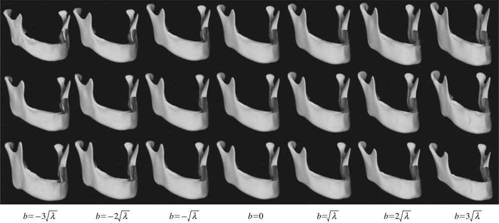
Fig. 4 Characteristic models of variation produced by PCA for male mandibles. The mode corresponding to the largest variance (λ1) is varied from to -3√λ1 to 3√λ1 (the first row), and the modes corresponding to the second mode (the second row) and the third mode (the third row).

Fig. 5 Characteristic models of variation produced by PCA for female mandibles. The mode corresponding to the largest variance (λ1) is varied from to -3√λ1 to 3√λ1 (the first row), and the modes corresponding to the second mode (the second row) and the third mode (the third row)

Fig. 6 Comparison of ramus lengths from the developed models with Ahn et al17 and Park et al.18

Fig. 7 Comparison of mandibular body lengths from the developed models with Ahn et al,17 Park et al,18 and Kim et al.19

Fig. 8 Comparison of gonial angles from the developed models with Ahn et al17 and Park et al.18
Reference
-
1. Franca C, Levin-Plotnik D, Sehgal V, Chen GT, Ramsey RG. Use of three-dimensional spiral computed tomography imaging for staging and surgical planning of head and neck cancer. J Digit Imaging. 2000; 13:24–32.
Article2. Elolf E, Tatagiba M, Samii M. Three-dimensional computed tomographic reconstruction: planning tool for surgery of skull base pathologies. Comput Aided Surg. 1998; 3:89–94.3. Troulis MJ, Everett P, Seldin EB, Kikinis R, Kaban LB. Development of a three-dimensional treatment planning system based on computer tomographic data. Int J Oral Maxillofac Surg. 2002; 31:349–357.4. Kragskov J, Sindet-Pedersen S, Gyldensted C, Jensen KL. A comparison of three-dimensional computed tomography scans and stereolithographic models for evaluation of craniofacial anomalies. J Oral Maxillofac Surg. 1996; 54:402–412.
Article5. Cheng AC, Wee AG. Reconstruction of cranial bone defects using alloplastic implants produced from a stereolithographically-generated cranial model. Ann Acad Med Singapore. 1999; 28:692–696.
Article6. Kermer C, Lindner A, Friede I, Wagner A, Millesi W. Preoperative stereolithographic model planning for primary reconstruction in craniomaxillofacial trauma surgery. J Craniomaxillofac Surg. 1998; 26:136–139.
Article7. Zachow S, Lamecker H, Elsholtz B, Stiller M. Reconstruction of mandibular dysplasia using a statistical 3D shape model. Int Congr Ser. 2005; 1281:1238–1243.
Article8. Gateno J, Xia JJ, Teichgraeber JF, Christensen AM, Lemoine JJ, Liebschner MA, et al. Clinical feasibility of computer-aided surgical simulation (CASS) in the treatment of complex craniomaxillofacial deformities. J Oral Maxillofac Surg. 2007; 65:728–734.9. D'Urso PS, Barker TM, Earwaker WJ, Bruce LJ, Atkinson RL, Lanigan MW, et al. Stereolithographic biomodelling in craniomaxillofacial surgery: a prospective trial. J Craniomaxillofac Surg. 1999; 27:30–37.10. Mavili ME, Canter HI, Saglam-Aydinatay B, Kamaci S, Kocadereli I. Use of three-dimensional medical modeling methods for precise planning of orthognathic surgery. J Craniofac Surg. 2007; 18:740–747.11. Hwang HS, Hwang CH, Lee KH, Kang BC. Maxillofacial 3-dimensional image analysis for the diagnosis of facial asymmetry. Am J Orthod Dentofacial Orthop. 2006; 130:779–785.12. Xia J, Ip HH, Samman N, Wang D, Kot CS, Yeung RW, et al. Computer-assisted three-dimensional surgical planning and simulation: 3D virtual osteotomy. Int J Oral Maxillofac Surg. 2000; 29:11–17.
Article13. Xia J, Samman N, Yeung RW, Shen SG, Wang D, Ip HH, et al. Three-dimensional virtual reality surgical planning and simulation workbench for orthognathic surgery. Int J Adult Orthodon Orthognath Surg. 2000; 15:265–282.
Article14. Cevidanes LH, Bailey LJ, Tucker GR Jr, Styner MA, Mol A, Phillips CL, et al. Superimposition of 3D cone-beam CT models of orthognathic surgery patients. Dentomaxillofac Radiol. 2005; 34:369–375.15. Katsumata A, Fujishita M, Maeda M, Ariji Y, Ariji E, Langlais RP. 3D-CT evaluation of facial asymmetry. Oral Surg Oral Med Oral Pathol Oral Radiol Endod. 2005; 99:212–220.16. Lamecker H, Zachow S, Wittmers A, Weber B, Hege HC, Elsholtz B, et al. Automatic segmentation of mandibles in lowdose CT data. Int J Comput Assist Radiol Surg. 2006; 1:suppl 1. 393–395.
Article17. Ahn JS, Lee KH, Hwang HS. A study on the 3-D standard value of mandible for the diagnosis of facial asymmetry. Korean J Orthod. 2005; 35:91–105.
Article18. Park SH, Yu HS, Kim KD, Baik HS. A proposal for a new analysis of craniofacial morphology by 3-dimensional computed tomography. Am J Orthod Dentofacial Orthop. 2006; 129:600.e23–600.e34.
Article19. Kim KH, Choy KC, Kim HG, Park KH. Cephalometric norms of the hard tissues of Korean for orthognatic surgery. J Korean Assoc Oral Maxillofac Surg. 2001; 27:221–230.
Article20. Brief J, Hassfeld S, Dauber S, Pernozzoli A, Munchenberg J, Redlich T, et al. 3D norm data: the first step towards semiautomatic virtual craniofacial surgery. Comput Aided Surg. 2000; 5:353–358.21. Eufinger H, Wehmoller M, Machtens E, Heuser L, Harders A, Kruse D. Reconstruction of craniofacial bone defects with individual alloplastic implants based on CAD/CAM-manipulated CT-data. J Craniomaxillofac Surg. 1995; 23:175–181.22. Brooks SL. Computed tomography. Dent Clin North Am. 1993; 37:575–590.
Article23. Cavalcanti MG, Haller JW, Vannier MW. Three-dimensional computed tomography landmark measurement in craniofacial surgical planning: experimental validation in vitro. J Oral Maxillofac Surg. 1999; 57:690–694.
Article
- Full Text Links
-
- Actions
-
Cited
- CITED
-
- Close
- Share
-
- Similar articles
-
- The Reliability of 3D CT Analysis for Facial Asymmetry
- A study on the cephalometric changes by the displacement of the mandibular condyles
- Cephalometric Angular Measurements of the Mandible Using Three-Dimensional Computed Tomography Scans in Koreans
- Comparison of measurements from digital cephalometric radiographs and 3D MDCT-synthetized cephalometric radiographs and the effect of head position
- Comparative study of glenoid version and inclination using two-dimensional images from computed tomography and three-dimensional reconstructed bone models
