Clinicoradiologic evaluation of styloid process calcification
- Affiliations
-
- 1Department of Oral Medicine Diagnosis and Radiology, M.N. D.A.V. Dental College and Hospital, Solan, India. munbhawni@gmail.com
- 2Department of Oral Medicine Diagnosis and Radiology, KD Dental College and Hospital, Mathura, India.
- KMID: 2167433
- DOI: http://doi.org/10.5624/isd.2012.42.3.155
Abstract
- PURPOSE
This study was performed to investigate the prevalence, morphology, and calcification pattern of the elongated styloid process in the Mathura population and its relation to gender, age, and mandibular movements.
MATERIALS AND METHODS
The study analyzed digital panoramic radiographs of 2,706 adults. The elongated styloid process was classified with the radiographic appearance based on the morphology and calcification pattern. The limits of mandibular protrusion were evaluated for each subject. The data were analyzed by using a Student's t-test and chi-squared test with significance set at p=0.05.
RESULTS
Bilateral elongation having an "elongated" type styloid process with a "partially mineralized" pattern was the most frequent type of styloid process. No correlation was found between styloid process type and calcification pattern on the one hand and gender on the other, although elongated styloid was more prevalent in older and male populations (p<0.05). Further styloid process elongation showed no effect on mandibular protrusive movement (p>0.05).
CONCLUSION
Dentists should recognize the existence of morphological variation in elongated styloid process or Eagle syndrome apparent on panoramic radiographs. We found higher prevalence of elongated styloid process in the population of the Mathura region when compared with other Indian populations. The calcification of the styloid process was more common in the older age group with no correlation to gender, mandibular movement and site. "Type I" with a "partially calcified" styloid process was observed more frequently in the population studied.
MeSH Terms
Figure
-
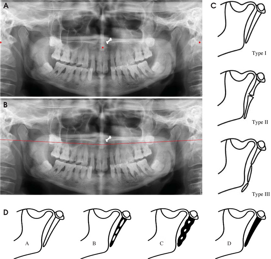
Fig. 1 A. The points show the anterior nasal spine and mastoid process on panoramic radiograph. B. Imaginary line connects the anterior nasal spine and the mastoid process on panoramic radiograph. C. Classification of elongation of styloid process based on type of calcification: Type I (elongated), Type II (pseudo articulated), and Type III (segmented). D. Classification of elongation of styloid process based on pattern of calcification (A: Calcified outline; B: Partially calcified; C: Nodular; D: Completely calcified).

Fig. 2 The diagram shows the number and percentage of the distribution of styloid process elongation according to the groups.
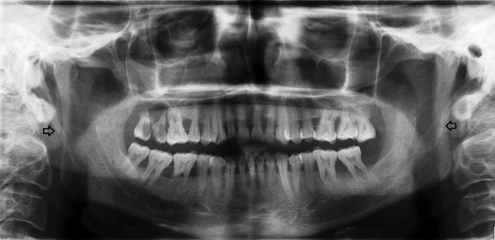
Fig. 3 A panoramic radiograph reveals bilateral styloid process calcification (Type I and Pattern B).
Cited by 1 articles
-
Nonsurgical treatment of stylohyoid (Eagle) syndrome: a case report
Arman Taheri, Shahram Firouzi-Marani, Masoud Khoshbin
J Korean Assoc Oral Maxillofac Surg. 2014;40(5):246-249. doi: 10.5125/jkaoms.2014.40.5.246.
Reference
-
1. Moffat DA, Ramsden RT, Shaw HJ. The styloid process syndrome: aetiological factors and surgical management. J Laryngol Otol. 1977. 91:279–294.
Article2. Feldman VB. Eagle's syndrome: a case of symptomatic calcification of the stylohyoid ligaments. J Can Chiropr Assoc. 2003. 47:21–27.3. Eagle WW. Elongated styloid processes. Report of two cases. Arch Otolaryngol Head Neck Surg. 1937. 25:548–587.
Article4. Keur JJ, Campbell JP, McCarthy JF, Ralph WJ. The clinical significance of the elongated styloid process. Oral Surg Oral Med Oral Pathol. 1986. 61:399–404.
Article5. Kursoglu P, Unalan F, Erdem T. Radiological evaluation of the styloid process in young adults resident in Turkey's Yeditepe University faculty of dentistry. Oral Surg Oral Med Oral Pathol Oral Radiol Endod. 2005. 100:491–494.
Article6. Ferrario VF, Sigurta D, Daddona A, Dalloca L, Miani A, Tafuro F, et al. Calcification of the stylohyoid ligament: incidence and morphoquantitative evaluations. Oral Surg Oral Med Oral Pathol. 1990. 69:524–529.
Article7. Langlais RP, Miles DA, Van Dis ML. Elongated and mineralized stylohyoid ligament complex: a proposed classification and report of a case of Eagle's syndrome. Oral Surg Oral Med Oral Pathol. 1986. 61:527–532.
Article8. Chouvel P, Rombaux P, Philips C, Hamoir M. Stylohyoid chain ossification: choice of the surgical approach. Acta Otorhinolaryngol Belg. 1996. 50:57–61.9. Gozil R, Yener N, Calguner E, Arac M, Tunc E, Bahcelioglu M. Morphological characteristics of styloid process evaluated by computerized axial tomography. Ann Anat. 2001. 183:527–535.10. Jung T, Tschernitschek H, Hippen H, Schneider B, Borchers L. Elongated styloid process: when is it really elongated? Dentomaxillofac Radiol. 2004. 33:119–124.
Article11. Savranlar A, Uzun L, Uğgur MB, Ozer T. Three-dimensional CT of Eagle's syndrome. Diagn Interv Radiol. 2005. 11:206–209.12. Ceylan A, Koybasioglu A, Celenk F, Yilmaz O, Uslu S. Surgical treatment of elongated styloid process: experience of 61 cases. Skull Base. 2008. 18:289–295.
Article13. More CB, Asrani MK. Evaluation of the styloid process on digital panoramic radiographs. Indian J Radiol Imaging. 2010. 20:261–265.
Article14. Gokce C, Sisman Y, Ertas ET, Akgunlu F, Ozturk A. Prevalence of styloid process elongation on panoramic radiography in the Turkey population from cappadocia region. Eur J Dent. 2008. 2:18–22.
Article15. Bozkir MG, Boga H, Dere F. The evaluation of elongated styloid process in panoramic radiographs in edentulous patients. Turk J Med Sci. 1999. 29:481–485.16. Phulambrikar T, Rajeshwari A, Rao BB, Warhekar A, Reddy P. Incidence of elongated styloid process: a radiographic study. J Indian Acad Oral Med Radiol. 2011. 23:S344–S346.
Article17. Yadav SP, Chanda R, Gera A, Yadav RK. Stylalgia: an Indian perspective. J Otolaryngol. 2001. 30:304–306.
Article18. Crowley LV. An introduction to human diseases: pathology and pathophysiology correlations. 2009. 8th ed. Sudbury: Jones and Bartlett Publishers;719–720.19. Srivastav A, Saraswat PK, Agarwal SK, Gupta P. A study of wrist ossification for age estimation in pediatric group in central Rajasthan. J Indian Acad Forensic Med. 2004. 26:132–135.20. Ilgüy M, Ilgüy D, Güler N, Bayirli G. Incidence of the type and calcification patterns in patients with elongated styloid process. J Int Med Res. 2005. 33:96–102.
Article21. Omnell KA, Gandhi C, Omnell ML. Ossification of the human stylohyoid ligament: a longitudinal study. Oral Surg Oral Med Oral Pathol Oral Radiol Endod. 1998. 85:226–232.22. Fini G, Gasparini G, Filippini F, Becelli R, Marcotullio D. The long styloid process syndrome or Eagle's syndrome. J Craniomaxillofac Surg. 2000. 28:123–127.
Article23. Camarda AJ, Deschamps C, Forest D. I. Stylohyoid chain ossification: a discussion of etiology. Oral Surg Oral Med Oral Pathol. 1989. 67:508–514.
Article24. Satyapal KS, Kalideen JM. Bilateral styloid chain ossification: case report. Surg Radiol Anat. 2000. 22:211–212.
Article25. Correll RW, Jensen JL, Taylor JB, Rhyne RR. Mineralization of the stylohyoid-stylomandibular ligament complex. A radiographic incidence study. Oral Surg Oral Med Oral Pathol. 1979. 48:286–291.26. Zaki HS, Greco CM, Rudy TE, Kubinski JA. Elongated styloid process in a temporomandibular disorder sample: prevalence and treatment outcome. J Prosthet Dent. 1996. 75:399–405.
Article
- Full Text Links
-
- Actions
-
Cited
- CITED
-
- Close
- Share
-
- Similar articles
-
- Radiologic Evaluation of Styloid Process by Fuchs' Series
- Severe calcified stylohyoid complex in twins: a case report
- Morphological study of styloid process of the temporal bone and its clinical implications
- A study on the styloid process in panoramic radiographs
- Measurement of Normal Size of Styloid Process with 3D Reconstruction CT
