Dedifferentiated Liposarcoma of the Retroperitoneum
- Affiliations
-
- 1Department of Urology, Bundang Jesaeng General Hospital, Seongnam, Korea. kkongpino@naver.com
Abstract
- A dedifferentiated liposarcoma of the retroperitoneum is an extremely rare tumor. A 51-year old man was admitted to our department because a retroperitoneal mass was seen on abdominal computed tomography at another hospital. Computed tomography of the abdomen and magnetic resonance imaging showed a large pelvic mass located in the right hemipelvis, and it was pushing the right ureter and invading the right kidney, duodenum, colon and inferior vena cava. The patient underwent right radical nephrectomy, pylorus preserving pancreatoduodenectomy, right hemicolectomy and artificial blood vessel replacement for the inferior vena cava. The histopathological diagnosis was dedifferentiated liposarcoma and the patient was free from recurrence on the computed tomography that was done 6 months after the operation.
Keyword
MeSH Terms
Figure
-
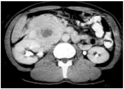
Fig. 1 CT of the abdomen shows a huge fat containing mass in the retroperitoneum. The right kidney, duodenum, inferior vena cava and pancreas are adhered to the mass.
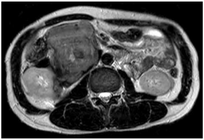
Fig. 2 MRI of the abdomen shows a huge solid soft tissue mass.

Fig. 3 On sectioning, the tumor mass shows a poorly circumscribed solid cut surface with multifocal necrosis, calcification and discrete intratumoral nodules of varying sizes. The right kidney is partially encased and the mass has grossly infiltrated the surrounding duodenum (thick arrow), the inferior vena cava wall (thin arrow), the pancreas and the colon.
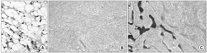
Fig. 4 The tumor consisted of a few components. (A) Well-differentiated liposarcoma area (H&E, ×400), (B) a fibrosarcomatous area (H&E, ×200) and (C) myxoid and neural whirling-like spindle cell nodules with multifocal metaplastic ossification (H&E, ×200).

Fig. 5 The follow-up CT taken six months after the operation shows no evidence of recurrence.
Reference
-
1. Antinori A, Antonacci V, Magistrelli P. Giant retroperitoneal liposarcoma. Am J Surg. 2002; 184:56–57. PMID: 12135721.
Article2. Moon WS, Jeong MJ, Lee DG, Choi HY, Kim SH. Dedifferentiated liposarcoma of the retroperitoneum: a case report. Korean J Pathol. 1993; 27:296–298.3. Cho YJ, Chun HJ, Park DK, Kim YB, Koh DW, Choung RS, et al. A case of dedifferentiated liposarcoma of retroperitoneum. Korean J Med. 2002; 62:552–556.4. Evans HL. Liposarcomas: a study of 55 cases with a reassessment of its classification. Am J Surg Pathol. 1979; 3:507–523. PMID: 534388.5. Tateishi U, Hasegawa T, Beppu Y, Satake M, Moriyama N. Primary dedifferentiated liposarcoma of the retroperitoneum. Prognostic significance of computed tomography and magnetic resonance imaging features. J Comput Assist Tomogr. 2003; 27:799–804. PMID: 14501373.6. Malkowicz SB. Retroperitoneal tumors: diagnosis, staging, surgery, management, and prognosis. Urologic oncology. 1997. 1st ed. Philadelphia: Saunders;p. 539–557.
Article7. Henricks WH, Chu YC, Goldblum JR, Weiss SW. Dedifferentiated liposarcoma: a clinicopathologic analysis of 155 cases with a proposal for an expanded definition of dedifferentiation. Am J Surg Pathol. 1997; 21:271–281. PMID: 9060596.
- Full Text Links
-
- Actions
-
Cited
- CITED
-
- Close
- Share
-
- Similar articles
-
- A Case of Dedifferentiated LiposarcomaThat Developed in the Dermis
- Dedifferentiated Liposarcoma of the Retroperitoneum: A case report
- Dedifferentiated Liposarcoma in the Thigh: Case Report
- A case of dedifferentiated liposarcoma in retroperitoneum
- Dedifferentiated Liposarcoma with a Peculiar Whorling Pattern: A Case Report
