Ann Rehabil Med.
2013 Apr;37(2):215-220. 10.5535/arm.2013.37.2.215.
Ultrasonographic Evaluation of Needle Insertion Site for the Flexor Pollicis Longus
- Affiliations
-
- 1Department of Rehabilitation Medicine, VHS Medical Center, Seoul, Korea. kkh702@korea.ac.kr
- KMID: 2165773
- DOI: http://doi.org/10.5535/arm.2013.37.2.215
Abstract
OBJECTIVE
To establish the safest approach to needle electrode insertion into the flexor pollicis longus (FPL) regarding possible needle injury to the superficial radial nerve (SRN) or radial artery by ultrasonography.
METHODS
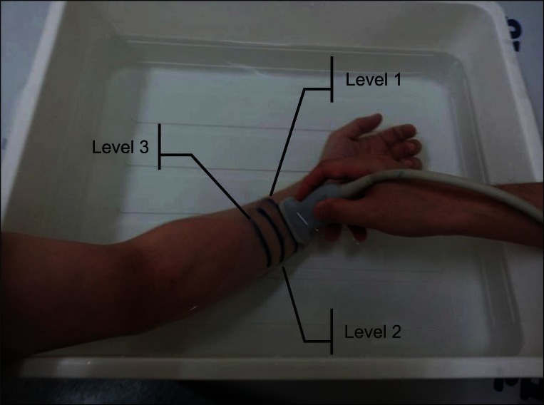
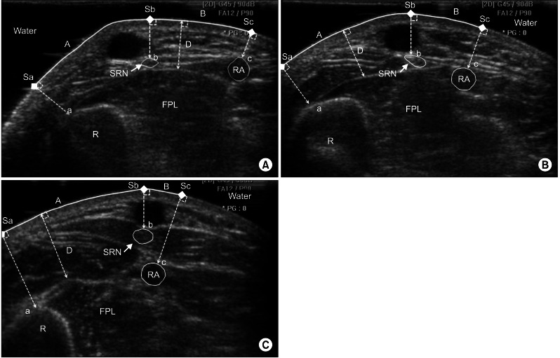
We evaluated 54 forearms of 27 healthy subjects. Three levels were defined in the forearm. Level 1 is the junction of the middle and distal third of the forearm, level 3 is the midpoint of forearm length, and level 2 is the midpoint between two levels. At each level, the distance between the most prominent point of the radius and the SRN (region A), the distance between the SRN and the radial artery (region B), and the depth from the skin surface to the FPL were measured.
RESULTS
The distance of region A was 1.20+/-0.41 cm in level 1, 1.62+/-0.45 cm in level 2, and 1.95+/-0.49 cm in level 3. The distance of region B was 1.02+/-0.29 cm in level 1, 0.61+/-0.24 cm in level 2, and 0.37+/-0.19 cm in level 3. The depth from the skin surface to the FPL was 0.92+/-0.20 cm in level 1, 1.14+/-0.26 cm in level 2, and 1.45+/-0.29 cm in level 3.
CONCLUSION
The safest needle insertion point to the FPL is the middle of the forearm within approximately 0.8 cm from the most prominent point of the radius. We recommend that the needle is inserted at the above point perpendicular to the skin surface until the needle meets the FPL at a depth of approximately 1.45 cm from the skin surface.
Keyword
MeSH Terms
Figure
Cited by 2 articles
-
Anatomical Basis of Pronator Teres for Electromyography Needle Placement Using Ultrasonography
Myung Kyu Park, In Yae Cheong, Ki Hoon Kim, Byung Kyu Park, Dong Hwee Kim
Ann Rehabil Med. 2015;39(1):39-46. doi: 10.5535/arm.2015.39.1.39.Optimal Radial Motor Nerve Conduction Study Using Ultrasound in Healthy Adults
Jungho Yeo, Yuntae Kim, Sooa Kim, Kiyoung Oh, Hyungdong Kang
Ann Rehabil Med. 2017;41(2):290-298. doi: 10.5535/arm.2017.41.2.290.
Reference
-
1. Jenkins DB. Hollinshead's functional anatomy of the limbs and back. 2009. 9th ed. Philadelphia: Saunders.2. Seror P. Anterior interosseous nerve lesions: clinical and electrophysiological features. J Bone Joint Surg Br. 1996; 78:238–241. PMID: 8666633.3. Pathak MS, Nguyen HT, Graham HK, Moore AP. Management of spasticity in adults: practical application of botulinum toxin. Eur J Neurol. 2006; 13(Suppl 1):42–50. PMID: 16417597.
Article4. Lee HJ, DeLisa JA. Manual of nerve conduction study and surface anatomy for needle electromyography. 2004. 4th ed. Philadelphia: Lippincott Williams & Wilkins.5. Perotto AO. Anatomical guide for the electromyographer: the limbs and trunk. 2005. 4th ed. Springfield: Charles C Thomas.6. Al-Shekhlee A, Shapiro BE, Preston DC. Iatrogenic complications and risks of nerve conduction studies and needle electromyography. Muscle Nerve. 2003; 27:517–526. PMID: 12707972.
Article7. Lee HJ, Bach JR, DeLisa JA. Needle electrode insertion into tibialis posterior: a new approach. Am J Phys Med Rehabil. 1990; 69:126–127. PMID: 2163649.8. Yang SN, Lee SH, Kwon HK. Needle electrode insertion into the tibialis posterior: a comparison of the anterior and posterior approaches. Arch Phys Med Rehabil. 2008; 89:1816–1818. PMID: 18760169.
Article9. Rha DW, Im SH, Lee SC, Kim SK. Needle insertion into the tibialis posterior: ultrasonographic evaluation of an anterior approach. Arch Phys Med Rehabil. 2010; 91:283–287. PMID: 20159135.
Article10. Won SJ, Kim JY, Yoon JS, Kim SJ. Ultrasonographic evaluation of needle electromyography insertion into the tibialis posterior using a posterior approach. Arch Phys Med Rehabil. 2011; 92:1921–1923. PMID: 21839985.
Article11. Boon AJ, Alsharif KI, Harper CM, Smith J. Ultrasound-guided needle EMG of the diaphragm: technique description and case report. Muscle Nerve. 2008; 38:1623–1626. PMID: 19016552.
Article12. Willenborg MJ, Shilt JS, Smith BP, Estrada RL, Castle JA, Koman LA. Technique for iliopsoas ultrasound-guided active electromyography-directed botulinum a toxin injection in cerebral palsy. J Pediatr Orthop. 2002; 22:165–168. PMID: 11856922.
Article13. Jordan SE, Ahn SS, Gelabert HA. Combining ultrasonography and electromyography for botulinum chemodenervation treatment of thoracic outlet syndrome: comparison with fluoroscopy and electromyography guidance. Pain Physician. 2007; 10:541–546. PMID: 17660852.14. Bechtold S, Rauch F, Noelle V, Donhauser S, Neu CM, Schoenau E, et al. Musculoskeletal analyses of the forearm in young women with Turner syndrome: a study using peripheral quantitative computed tomography. J Clin Endocrinol Metab. 2001; 86:5819–5823. PMID: 11739445.
Article15. Neu CM, Rauch F, Rittweger J, Manz F, Schoenau E. Influence of puberty on muscle development at the forearm. Am J Physiol Endocrinol Metab. 2002; 283:E103–E107. PMID: 12067849.
Article16. Blaivas M, Lyon M, Brannam L, Duggal S, Sierzenski P. Water bath evaluation technique for emergency ultrasound of painful superficial structures. Am J Emerg Med. 2004; 22:589–593. PMID: 15666267.
Article17. Dumitru D, Amato AA, Zwarts M. Electrodiagnostic medicine. 1995. 2nd ed. Philadelphia: Hanley & Belfus.
- Full Text Links
- Actions
-
Cited
- CITED
-
- Close
- Share
- Similar articles
-
- Ultrasonographic Evaluation of the Needle Insertion Site for the Flexor Pollicis Longus Using the Flexor Carpi Radialis Tendon
- Flexor Pollicis Longus Tendon Rupture due to Scaphoid Nonunion
- Flexor Pollicis Longus Reconstruction in Patient with the Linburg-Comstock Syndrome
- Pathologic Rupture of Flexor Pollicis Longus Tendon Secondaryto Kienbock's Disease: A Case Report
- Variations of Insertions of the Abductor Pollicis Longus and the Extensor Pollicis Brevis in Korean


