Outcomes of Ultrasound-Guided Extracorporeal Shock Wave Therapy for Painful Stump Neuroma
- Affiliations
-
- 1Department of Rehabilitation Medicine, Dongtan Sacred Heart Hospital, Hallym University College of Medicine, Hwaseong, Korea.
- 2Department of Rehabilitation Medicine, Hangang Sacred-Heart Hospital, Hallym University College of Medicine, Seoul, Korea. s9036@naver.com
- KMID: 2165735
- DOI: http://doi.org/10.5535/arm.2014.38.4.523
Abstract
OBJECTIVE
To investigate the effect of extracorporeal shock wave therapy (ESWT) on painful stump neuroma.
METHODS
Thirty patients with stump neuroma at the distal end of an amputation site were assigned randomly to the ESWT group (n=15) and the transcutaneous electrical nerve stimulation (TENS)+desensitization+pharmacological treatment group (n=15). For 3 weeks, the ESWT group received a weekly session involving 1,500 pulses at 0.10 mJ/mm2, while the control group was treated 10 times each, 40 minutes per day with TENS and desensitization treatment, and daily medication for 3 weeks. ESWT stimulation was given by focusing on the area at the neuroma site clearly identified by ultrasound.
RESULTS
The changes in the McGill pain questionnaire were 38.8+/-9.0 prior to treatment and 11.8+/-3.1 following the treatment. The corresponding values for the control group were 37.2+/-7.7 and 28.5+/-10.3. The changes between groups were significantly different (p=0.035). The change in visual analog scale prior to and after treatment was 7.0+/-1.5 and 2.8+/-0.8 in the ESWT group, respectively, and 7.2+/-1.4 and 5.8+/-2.0 in the control group. These changes between the groups were also significantly different (p=0.010). The outcome in the pain rating scale also showed significant differences between groups (p<0.001). Changes in neuroma size and pain pressure threshold (lb/cm2) were not significantly different between groups (p>0.05).
CONCLUSION
The study findings imply that ESWT for stump neuroma is superior to conventional therapy.
Keyword
MeSH Terms
Figure
-
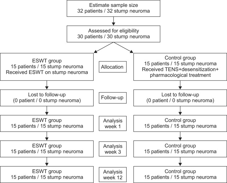
Fig. 1 Flow chart of extracorporeal shock wave therapy (ESWT) for stump neuroma. TENS, transcutaneous electrical nerve stimulation.
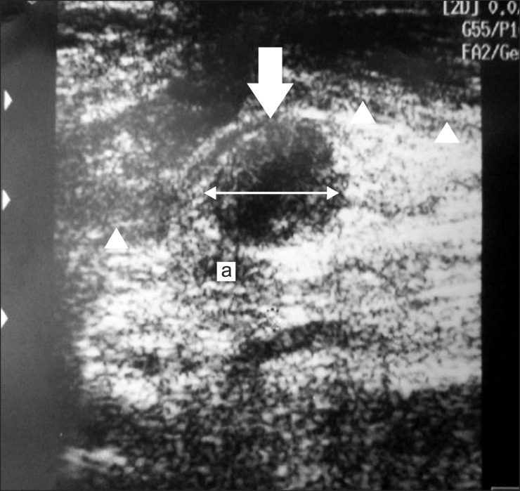
Fig. 2 Transverse ultrasound image of medial ankle reveals ovoid, hypoechoic mass (arrow), directly continuous to the posterior tibial nerve. Posterior tibial neuroma is close to the posterior tibial artery (a) and covered by flexor retinaculum (arrowhead). Digital calipers indicate the size of the neuroma to be 16.1 mm.
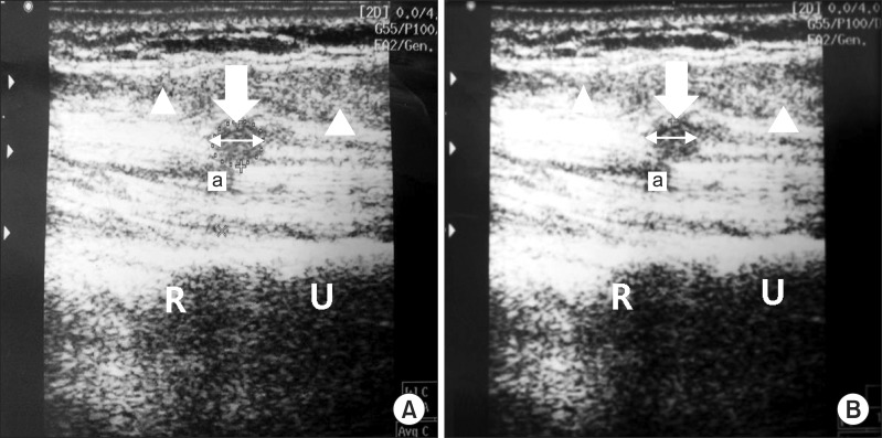
Fig. 3 A 43-year-old male (patient no. 7) with a transradial stump neuroma (arrow). Scanning over the proximal forearm demonstrates end stump of radius (R) and ulnar (U) bone. Transverse ultrasound image of proximal forearm showed hypoechoic mass (arrow) below the humeral head of pronator teres muscle (arrowhead) and close to the ulnar artery (a). No change of neuroma size was apparent before and following extracorporeal shock wave therapy (ESWT) treatment. (A) The neuroma of initial assessment before ESWT treatment. Digital calipers indicate the size of the neuroma is 10.6 mm. (B) The neuroma at the final assessment after ESWT treatment. Digital calipers indicate the size of the neuroma is 10.8 mm.
Reference
-
1. Provost N, Bonaldi VM, Sarazin L, Cho KH, Chhem RK. Amputation stump neuroma: ultrasound features. J Clin Ultrasound. 1997; 25:85–89. PMID: 9023697.
Article2. Henrot P, Stines J, Walter F, Martinet N, Paysant J, Blum A. Imaging of the painful lower limb stump. Radiographics. 2000; 20 Spec No:S219–S235. PMID: 11046173.
Article3. Ernberg LA, Adler RS, Lane J. Ultrasound in the detection and treatment of a painful stump neuroma. Skeletal Radiol. 2003; 32:306–309. PMID: 12719933.
Article4. Vernadakis AJ, Koch H, Mackinnon SE. Management of neuromas. Clin Plast Surg. 2003; 30:247–268. PMID: 12737355.
Article5. Whipple RR, Unsell RS. Treatment of painful neuromas. Orthop Clin North Am. 1988; 19:175–185. PMID: 3275925.
Article6. Thomas AJ, Bull MJ, Howard AC, Saleh M. Peri operative ultrasound guided needle localisation of amputation stump neuroma. Injury. 1999; 30:689–691. PMID: 10707244.
Article7. Liang HW, Wang TG, Chen WS, Hou SM. Thinner plantar fascia predicts decreased pain after extracorporeal shock wave therapy. Clin Orthop Relat Res. 2007; 460:219–225. PMID: 17353798.
Article8. Flor H. Phantom-limb pain: characteristics, causes, and treatment. Lancet Neurol. 2002; 1:182–189. PMID: 12849487.
Article9. Fisher GT, Boswick JA Jr. Neuroma formation following digital amputations. J Trauma. 1983; 23:136–142. PMID: 6827633.
Article10. Dockery GL. The treatment of intermetatarsal neuromas with 4% alcohol sclerosing injections. J Foot Ankle Surg. 1999; 38:403–408. PMID: 10614611.
Article11. Sobiesk GA, Wertheimer SJ, Schulz R, Dalfovo M. Sonographic evaluation of interdigital neuromas. J Foot Ankle Surg. 1997; 36:364–366. PMID: 9356915.
Article12. Wu J, Chiu DT. Painful neuromas: a review of treatment modalities. Ann Plast Surg. 1999; 43:661–667. PMID: 10597831.13. Speed CA. Extracorporeal shock-wave therapy in the management of chronic soft-tissue conditions. J Bone Joint Surg Br. 2004; 86:165–171. PMID: 15046427.
Article14. Jung KH, Hwang JH, Chang HJ, Yoon YC, Park MJ, Yoo JC, et al. Low-energy extracorporeal shock wave therapy on chronic epicondylitis of the elbow: clinical and sonographic study. J Korean Acad Rehabil Med. 2009; 33:77–83.15. Kim SB, Lee KW, Lee JH, Kim YD, Yoon K, Joe YL. The effect of extracorporeal shock wave therapy in plantar fasciitis. J Korean Acad Rehabil Med. 2009; 33:333–338.16. Fridman R, Cain JD, Weil L Jr. Extracorporeal shockwave therapy for interdigital neuroma: a randomized, placebo-controlled, double-blind trial. J Am Podiatr Med Assoc. 2009; 99:191–193. PMID: 19448168.17. Mathews GJ, Osterholm JL. Painful traumatic neuromas. Surg Clin North Am. 1972; 52:1313–1324. PMID: 4561764.
Article18. Hagenacker T, Ledwig D, Busselberg D. Feedback mechanisms in the regulation of intracellular calcium ([Ca2+]i) in the peripheral nociceptive system: role of TRPV-1 and pain related receptors. Cell Calcium. 2008; 43:215–227. PMID: 17673288.
Article19. Fioramonti P, Cigna E, Onesti MG, Fino P, Fallico N, Scuderi N. Extracorporeal shock wave therapy for the management of burn scars. Dermatol Surg. 2012; 38:778–782. PMID: 22335776.
Article
- Full Text Links
-
- Actions
-
Cited
- CITED
-
- Close
- Share
-
- Similar articles
-
- Extracorporeal Shock Wave Therapy For Treatment of Intractable Stump Pain
- Sonographically Guided Alcohol Injection in Painful Stump Neuroma
- Current Concepts in Extracorporeal Shock Wave Therapy
- Chronic Intractable Calcific Lateral Epicondylopathy Treated with Ultrasound-Guided Barbotage Combined with Extracorporeal Shock-Wave Therapy
- Comparison of Extracorporeal Shock Wave Therapy and Ultrasound-Guided Shoulder Injection Therapy in Patients with Supraspinatus Tendinitis
