Endoscopic Molecular Imaging: Status and Future Perspective
- Affiliations
-
- 1Department of Gastroenterology and Oncology, Institute of Health Biosciences, The University of Tokushima Graduate School, Tokushima, Japan. muguruma.clin.med@gmail.com
Abstract
- During the last decade, researchers have made great progress in the development of new image processing technologies for gastrointestinal endoscopy. However, diagnosis using conventional endoscopy with white light optical imaging is essentially limited, and ultimately, we still rely on the histopathological diagnosis from biopsy specimens. Molecular imaging represents the most novel imaging methods in medicine, and the future of endoscopic diagnosis is likely to be impacted by a combination of biomarkers and technology. Endoscopic molecular imaging can be defined as the visualization of molecular characteristics with endoscopy. These innovations will allow us not only to locate a tumor or dysplastic lesion but also to visualize its molecular characteristics and the activity of specific molecules and biological processes that affect tumor behavior and/or its response to therapy. In the near future, these promising technologies will play a central role in endoluminal oncology.
MeSH Terms
Figure
-
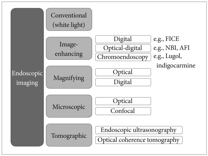
Fig. 1 Endoscopic imaging classification proposed by Tajiri and Niwa (modified by the authors). FICE, flexible spectral imaging color enhancement; NBI, narrow band imaging; AFI, autofluorescence imaging.

Fig. 2 Strategy based on the imaging classification in endoluminal oncology. ESD, endoscopic submucosal dissection; EMR, endoscopic mucosal resection; OCT, optical coherence tomography; EUS, endoscopic ultrasound.
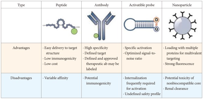
Fig. 3 Comparison of different molecular probe classes. Adapted from Goetz et al. Gastroenterology 2010;138:828-833, with permission from Elsevier.32 ab, antibody.
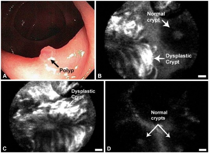
Fig. 4 In vivo confocal fluorescence images of the border between a colonic adenoma and normal mucosa showing peptide binding to dysplastic colonocytes. (A) Endoscopic view. (B) Border. (C) Dysplastic crypt. (D) Adjacest mucosa. Scale bars, 20 µm.
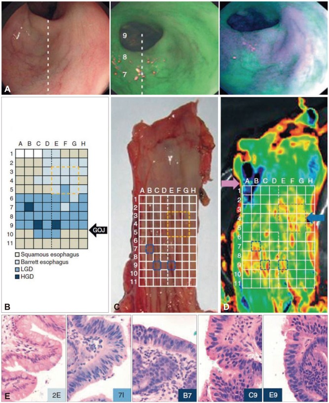
Fig.5 Molecular imaging of an esophagectomy specimen with a 6-cm segment of Barrett esophagus containing macroscopically invisible residual high grade dysplasia (HGD) and focal intramucosal carcinoma. (A) Images taken with an endoscope. White-light image (left), imaging fluorescence at 490 to 560 nm before white germ agglutinin. Application (middle) and imaging fluorescence at 490 to 560 nm after wheat germ agglutinin (WGA) and Alexa Fluor 488 application (right). The areas of low WGA binding appear in purple. (B) The dashed white line is placed longitudinally along the posterior wall of the esophagus to facilitate orientation between the different images, and the numbers 7, 8, and 9 refer to the y coordinates on the reference grid in. White-light imaging of the lower esophagus revealed no macroscopic abnormalities such as ulcers or nodules, and before WGA application, we detected no appreciable differences in mucosal autofluorescence; however, after incubation with WGA, differences in lectin binding were evident. High binding is represented by a green signal and low binding is represented by a purple signal on the pseudocolor image. Grid showing the pathological diagnostic map (color-coded, with darker colors representing a worsening grade of dysplasia) of each block made from the resection specimen. This same grid can be compared with the endoscopic and in vivo imaging system (IVIS) fluorescence images in (A), on the right, and in (D). The dashed line represents the longitudinal axis along the posterior wall of the esophagus. (C) The same specimen after being opened longitudinally along the anterior border of the esophagus is shown with the overlying grid from (B). (D) The WGA fluorescence signal from the esophageal specimen taken with the IVIS 200 camera. The pink arrow marks an area of artifact from the exposed submucosal tissue, and the blue arrow indicates the site of a previous endoscopic mucosal resection (outlined with a dashed gray box). The specimen was cut into 11 transverse sections (rows labeled 1 to 11), and the pathologist divided each of these further into 8 areas (columns labeled A-H) to allow mapping. (E) Examples of the histological appearance (×40) at various coordinates from the grid. From left to right, the images show nondysplastic Barrett esophagus, low grade dysplasia (LGD), and two examples of HGD. The corresponding grid reference is given at the bottom of each image. Adapted from Bird-Lieberman et al. Nat Med 2012;18:315-321, with permission from Nature Publishing Group.40
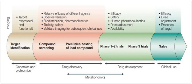
Fig. 6 Imaging applications in the drug discovery and development process. Adapted from Rudin et al. Nat Rev Drug Discov 2003;2:123-131, with permission from Nature Publishing Group.31
Reference
-
1. Sivak MV. Gastrointestinal endoscopy: past and future. Gut. 2006; 55:1061–1064. PMID: 16849338.
Article2. Cotton PB, Barkun A, Ginsberg G, et al. Diagnostic endoscopy: 2020 vision. Gastrointest Endosc. 2006; 64:395–398. PMID: 16923489.
Article3. Uedo N, Iishi H, Tatsuta M, et al. A novel videoendoscopy system by using autofluorescence and reflectance imaging for diagnosis of esophagogastric cancers. Gastrointest Endosc. 2005; 62:521–528. PMID: 16185965.
Article4. Gono K, Obi T, Yamaguchi M, et al. Appearance of enhanced tissue features in narrow-band endoscopic imaging. J Biomed Opt. 2004; 9:568–577. PMID: 15189095.
Article5. Jung SW, Lim KS, Lim JU, et al. Flexible spectral imaging color enhancement (FICE) is useful to discriminate among non-neoplastic lesion, adenoma, and cancer of stomach. Dig Dis Sci. 2011; 56:2879–2886. PMID: 21800158.
Article6. Hong SN, Choe WH, Lee JH, et al. Prospective, randomized, back-to-back trial evaluating the usefulness of i-SCAN in screening colonoscopy. Gastrointest Endosc. 2012; 75:1011–1021. PMID: 22381530.
Article7. Kiesslich R, Burg J, Vieth M, et al. Confocal laser endoscopy for diagnosing intraepithelial neoplasias and colorectal cancer in vivo. Gastroenterology. 2004; 127:706–713. PMID: 15362025.
Article8. Tajiri H, Niwa H. Proposal for a consensus terminology in endoscopy: how should different endoscopic imaging techniques be grouped and defined? Endoscopy. 2008; 40:775–778. PMID: 18698532.
Article9. Weissleder R, Mahmood U. Molecular imaging. Radiology. 2001; 219:316–333. PMID: 11323453.
Article10. Thakur M, Lentle BC. Report of a summit on molecular imaging. Radiology. 2005; 236:753–755. PMID: 16118158.
Article11. Mahmood U, Wallace MB. Molecular imaging in gastrointestinal disease. Gastroenterology. 2007; 132:11–14. PMID: 17241854.
Article12. Takayama T, Katsuki S, Takahashi Y, et al. Aberrant crypt foci of the colon as precursors of adenoma and cancer. N Engl J Med. 1998; 339:1277–1284. PMID: 9791143.
Article13. Keller R, Winde G, Eisenhawer C, et al. Immunoscopy: a technique combining endoscopy and immunofluorescence for diagnosis of colorectal carcinoma. Gastrointest Endosc. 1998; 47:154–161. PMID: 9512281.14. Pasricha PJ, Motamedi M. Optical biopsies, "bioendoscopy," and why the sky is blue: the coming revolution in gastrointestinal imaging. Gastroenterology. 2002; 122:571–575. PMID: 11832471.
Article15. Fujimoto JG, Brezinski ME, Tearney GJ, et al. Optical biopsy and imaging using optical coherence tomography. Nat Med. 1995; 1:970–972. PMID: 7585229.
Article16. Weissleder R. Molecular imaging in cancer. Science. 2006; 312:1168–1171. PMID: 16728630.
Article17. Goetz M, Hoetker MS, Diken M, Galle PR, Kiesslich R. In vivo molecular imaging with cetuximab, an anti-EGFR antibody, for prediction of response in xenograft models of human colorectal cancer. Endoscopy. 2013; 45:469–477. PMID: 23580409.
Article18. Mitsunaga M, Ogawa M, Kosaka N, Rosenblum LT, Choyke PL, Kobayashi H. Cancer cell-selective in vivo near infrared photoimmunotherapy targeting specific membrane molecules. Nat Med. 2011; 17:1685–1691. PMID: 22057348.
Article19. Keller R, Winde G, Terpe HJ, Foerster EC, Domschke W. Fluorescence endoscopy using a fluorescein-labeled monoclonal antibody against carcinoembryonic antigen in patients with colorectal carcinoma and adenoma. Endoscopy. 2002; 34:801–807. PMID: 12244502.
Article20. Marten K, Bremer C, Khazaie K, et al. Detection of dysplastic intestinal adenomas using enzyme-sensing molecular beacons in mice. Gastroenterology. 2002; 122:406–414. PMID: 11832455.
Article21. Hsiung PL, Hardy J, Friedland S, et al. Detection of colonic dysplasia in vivo using a targeted heptapeptide and confocal microendoscopy. Nat Med. 2008; 14:454–458. PMID: 18345013.
Article22. Ito S, Muguruma N, Kusaka Y, et al. Detection of human gastric cancer in resected specimens using a novel infrared fluorescent anti-human carcinoembryonic antigen antibody with an infrared fluorescence endoscope in vitro. Endoscopy. 2001; 33:849–853. PMID: 11571680.
Article23. Vogelstein B, Papadopoulos N, Velculescu VE, Zhou S, Diaz LA Jr, Kinzler KW. Cancer genome landscapes. Science. 2013; 339:1546–1558. PMID: 23539594.
Article24. Funovics MA, Alencar H, Montet X, Weissleder R, Mahmood U. Simultaneous fluorescence imaging of protease expression and vascularity during murine colonoscopy for colonic lesion characterization. Gastrointest Endosc. 2006; 64:589–597. PMID: 16996355.25. Petrovsky A, Schellenberger E, Josephson L, Weissleder R, Bogdanov A Jr. Near-infrared fluorescent imaging of tumor apoptosis. Cancer Res. 2003; 63:1936–1942. PMID: 12702586.26. Citrin D, Lee AK, Scott T, et al. In vivo tumor imaging in mice with near-infrared labeled endostatin. Mol Cancer Ther. 2004; 3:481–488. PMID: 15078992.27. Hoetker MS, Kiesslich R, Diken M, et al. Molecular in vivo imaging of gastric cancer in a human-murine xenograft model: targeting epidermal growth factor receptor. Gastrointest Endosc. 2012; 76:612–620. PMID: 22771099.
Article28. Goetz M, Ziebart A, Foersch S, et al. In vivo molecular imaging of colorectal cancer with confocal endomicroscopy by targeting epidermal growth factor receptor. Gastroenterology. 2010; 138:435–446. PMID: 19852961.
Article29. Foersch S, Kiesslich R, Waldner MJ, et al. Molecular imaging of VEGF in gastrointestinal cancer in vivo using confocal laser endomicroscopy. Gut. 2010; 59:1046–1055. PMID: 20639250.
Article30. Bando T, Muguruma N, Ito S, et al. Basic studies on a labeled anti-mucin antibody detectable by infrared-fluorescence endoscopy. J Gastroenterol. 2002; 37:260–269. PMID: 11993509.
Article31. Rudin M, Weissleder R. Molecular imaging in drug discovery and development. Nat Rev Drug Discov. 2003; 2:123–131. PMID: 12563303.
Article32. Goetz M, Wang TD. Molecular imaging in gastrointestinal endoscopy. Gastroenterology. 2010; 138:828–833. PMID: 20096697.
Article33. Kelly K, Alencar H, Funovics M, Mahmood U, Weissleder R. Detection of invasive colon cancer using a novel, targeted, library-derived fluorescent peptide. Cancer Res. 2004; 64:6247–6251. PMID: 15342411.
Article34. Fujikawa Y, Urano Y, Komatsu T, et al. Design and synthesis of highly sensitive fluorogenic substrates for glutathione S-transferase and application for activity imaging in living cells. J Am Chem Soc. 2008; 130:14533–14543. PMID: 18841967.
Article35. Joshi BP, Liu Z, Elahi SF, Appelman HD, Wang TD. Near-infrared-labeled peptide multimer functions as phage mimic for high affinity, specific targeting of colonic adenomas in vivo (with videos). Gastrointest Endosc. 2012; 76:1197–1206. PMID: 23022051.
Article36. Wu X, Liu H, Liu J, et al. Immunofluorescent labeling of cancer marker Her2 and other cellular targets with semiconductor quantum dots. Nat Biotechnol. 2003; 21:41–46. PMID: 12459735.
Article37. Mordon S, Devoisselle JM, Soulie-Begu S, Desmettre T. Indocyanine green: physicochemical factors affecting its fluorescence in vivo. Microvasc Res. 1998; 55:146–152. PMID: 9521889.38. Ogawa M, Kosaka N, Choyke PL, Kobayashi H. In vivo molecular imaging of cancer with a quenching near-infrared fluorescent probe using conjugates of monoclonal antibodies and indocyanine green. Cancer Res. 2009; 69:1268–1272. PMID: 19176373.
Article39. Nussbaum S, Roth HJ. Human anti-mouse antibodies: pitfalls in tumor marker measurement and strategies for enhanced assay robustness: including results with Elecsys CEA. Anticancer Res. 2000; 20(6D):5249–5252. PMID: 11326704.40. Bird-Lieberman EL, Neves AA, Lao-Sirieix P, et al. Molecular imaging using fluorescent lectins permits rapid endoscopic identification of dysplasia in Barrett's esophagus. Nat Med. 2012; 18:315–321. PMID: 22245781.
Article41. Ozdemir V, Williams-Jones B, Glatt SJ, Tsuang MT, Lohr JB, Reist C. Shifting emphasis from pharmacogenomics to theragnostics. Nat Biotechnol. 2006; 24:942–946. PMID: 16900136.
Article42. Weissleder R. Molecular imaging: exploring the next frontier. Radiology. 1999; 212:609–614. PMID: 10478223.
Article43. Barrett T, Koyama Y, Hama Y, et al. In vivo diagnosis of epidermal growth factor receptor expression using molecular imaging with a cocktail of optically labeled monoclonal antibodies. Clin Cancer Res. 2007; 13(22 Pt 1):6639–6648. PMID: 17982120.
- Full Text Links
-
- Actions
-
Cited
- CITED
-
- Close
- Share
-
- Similar articles
-
- Endoscopic molecular imaging in inflammatory bowel disease
- Nuclear Imaging of Differentiated Thyroid Cancer: Current Status and Future Perspective
- Deep Learning in Nuclear Medicine and Molecular Imaging: Current Perspectives and Future Directions
- General Perspectives for Molecular Nuclear Imaging
- Molecular MR Imaging
