Development of Biliary and Enteral Stents by the Korean Gastrointestinal Endoscopists
- Affiliations
-
- 1Digestive Disease Center, Konkuk University Medical Center, Konkuk University School of Medicine, Seoul, Korea. chansshim@kuh.ac.kr
- 2Department of Gastroenterology, Ajou University Hospital, Ajou University School of Medicine, Suwon, Korea.
- 3Department of Internal Medicine, Min Hospital, Seoul, Korea.
- KMID: 2165034
- DOI: http://doi.org/10.5946/ce.2016.039
Abstract
- Stenting in the gastrointestinal tract is a common procedure used for palliation of obstruction in the enteral and biliary tract. Today, stenting of malignant and benign strictures is performed at almost every major tertiary hospital in Korea. Moreover, Korea has become a major global supplier of cutting edge technology in the field of self-expanding metal stents. However, the history of stenting in Korea is relatively short and was far behind that of other nations such as Japan and Germany. The authors are humbled and gratified to have been able to observe the development and application of these stents in Korea, first hand. In this article, the authors review the overall history of stenting with a specific focus on the development of stenting in Korea. The development of esophageal, gastroduodenal, biliary, and colonic stents in Korea are reviewed in this article from a chronological and historical point of view, and a personal account of some of the significant moments of stent development in Korea are described.
MeSH Terms
Figure
-

Fig. 1. Hand-made plastic biliary stent with side holes and side flaps (10 mm in diameter).

Fig. 2. Endoscopic esophageal stenting in esophageal cancer with a celestin stent. (A) Celestin stent. (B) Pusher tube (*), stent (**). (C) Esophagogram of mid esophageal cancer. (D) Bougie dilation state. (E) Stent over the endoscope and pusher tube as an introducer clips for marking the length of tumor. (F) Stent just after deployment.

Fig. 3. Special esophageal stents for unusual situations. (A) Self-expandable metal stent (SEMS) with antireflux mechanism with long S-shaped flap. (B) SEMS for upper cervical esophageal cancer, a 7 mm short upper end of stent in a flask shape to prevent migration. (C) SEMS with Shim’s technique for antimigration.

Fig. 4. Hand-made membrane covered biliary self-expandable metal stent. Membrane-covered self-expandable biliary metal stent in expansion after uncoiling the string modified membrane-covered self-expandable biliary metal stent. Stent material, stainless steel 30 Fr; membrane, polyurethane; introducing apparatus, string pull type.

Fig. 5. Covered biliary self-expandable metal stent (SEMS). (A, B) Covered biliary SEMSs and delivery devices. (C-F) Covered biliary SEMSs and expansion from captured position. Implantation of biliary stent into a malignant biliary patient. (G) A X-ray image of fully deployed biliary stent, (H) endoscopic images of fully deployed biliary stent.
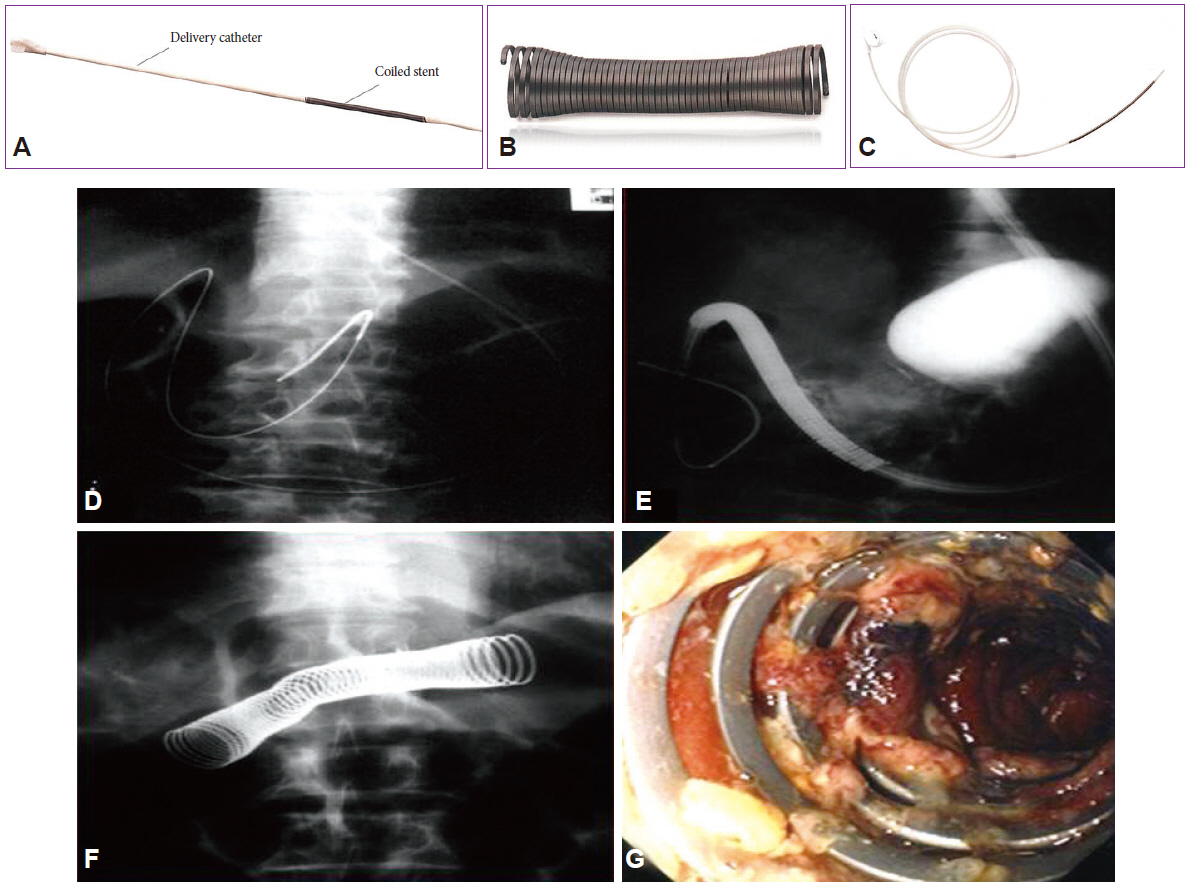
Fig. 6. (A) Original delivery catheter and coiled stent for esophagal stenting. (B) Deployed EsophaCoil (InStent) in vitro. (C) Modified and extended delivery catheter to 150 cm in length and mounted stent for antroduodenal stenting. Implanting a metallic stent (EsophaCoil stent; InStent) under the fluoroscopic guidance. (D) An image of guide wire inserted deep into duodenum using fluoroscopic image. (E) An image of EsophaCoil stent being inserted following a guide wire. (F) X-ray image after the deployment completed. (G) An endoscopic image of metallic stent placed in gastric outlet region.

Fig. 7. First trial of rectal plastic stent in 1988. (A) An esophageal plastic stent (Sumitomo-Bakelite Stent; Sumitomo-Bakelite). (B) X-ray image of an esophageal plastic stent in a malignant obstructive rectal cancer patient (an esophageal stent was placed in reverse).

Fig. 8. First trial colonic through-the-scope (TTS) self-expandable metal stent. (A) The partly membrane covered Niti-S stent (Taewoong Medical) with flanges. The proximal part of the flange is not covered with polyurethane. (B) The uncovered Niti-S colon stent, without flanges. (C) The stent-in-stent model (with an outer uncovered stent and an inner partly covered Niti-S stent). (D) Easy stent placement being practiced under an endoscopic view at any obstructed region. Large diameter TTS colonic stent are easily being inserted into a colonoscope channel for easier monitoring of expansion.
Reference
-
1. Shim CS, Yoon SJ, Paik SH, et al. Case series of endoscopic retrograde biliary drainage (ERBD) in malignant obstructive jaundice. Korean J Gastrointest Endosc. 1986; 6(Suppl):88–89.2. Celestin LR. Permanent intubation in inoperable cancer of the oesophagus and cardia: a new tube. Ann R Coll Surg Engl. 1959; 25:165–170.3. Duseja A, Chawla YK, Singh RP, Sharma TR, Kaur U, Dilawari JB. Dilatation of benign oesophageal strictures: 10 years’ experience with Celestin dilators. J Gastroenterol Hepatol. 2000; 15:26–29.
Article4. Tytgat GN, den Hartog Jager FC. Non-surgical treatment of cardio-esophageal obstruction: role of endoscopy. Endoscopy. 1977; 9:211–215.5. Hussain SA, Hughes R, Gross E. Dangers from dissolution of latex in Celestin endo-oesophageal tube. Br Med J (Clin Res Ed). 1987; 294:412–413.
Article6. Shim CS, Cho SW, Kim JH, Cho SW. Experience with endoscopic esophageal endoprosthesis in malignant esophagogastric strictures. Korean J Med. 1989; 36:507–516.7. Olsen E, Thyregaard R, Kill J. Esophacoil expanding stent in the management of patients with nonresectable malignant esophageal or cardiac neoplasm: a prospective study. Endoscopy. 1999; 31:417–420.8. Song HY, Choi KC, Cho BH, Ahn DS, Kim KS. Esophagogastric neoplasms: palliation with a modified gianturco stent. Radiology. 1991; 180:349–354.
Article9. Park HS, Do YS, Suh SW, et al. Upper gastrointestinal tract malignant obstruction: initial results of palliation with a flexible covered stent. Radiology. 1999; 210:865–870.
Article10. Do YS, Choo SW, Suh SW, et al. Malignant esophagogastric junction obstruction: palliative treatment with an antireflux valve stent. J Vasc Interv Radiol. 2001; 12:647–651.11. Kim MD, Park SB, Kang DH, et al. Double layered self-expanding metal stents for malignant esophageal obstruction, especially across the gastroesophageal junction. World J Gastroenterol. 2012; 18:3732–3737.
Article12. Shim CS, Jung IS, Cheon YK, et al. Management of malignant stricture of the esophagogastric junction with a newly designed self-expanding metal stent with an antireflux mechanism. Endoscopy. 2005; 37:335–339.
Article13. Shim CS, Jung IS, Bhandari S, et al. Management of malignant strictures of the cervical esophagus with a newly-designed self-expanding metal stent. Endoscopy. 2004; 36:554–557.
Article14. Shim CS, Cho YD, Moon JH, et al. Fixation of a modified covered esophageal stent: its clinical usefulness for preventing stent migration. Endoscopy. 2001; 33:843–848.
Article15. Jeon SR, Eun SH, Shim CS, et al. Effect of drug-eluting metal stents in benign esophageal stricture: an in vivo animal study. Endoscopy. 2009; 41:449–456.
Article16. Lee KM, Shin SJ, Hwang JC, et al. Proximal-releasing stent insertion under transnasal endoscopic guidance in patients with postoperative esophageal leakage. Gastrointest Endosc. 2010; 72:180–185.
Article17. Cheon YK, Lee TY, Sung IK, Shim CS. Clinical feasibility of a new through-the-scope fully covered esophageal self-expandable metallic stent: an in vivo animal study. Dig Endosc. 2014; 26:32–36.18. Soehendra N, Reynders-Frederix V. Palliative biliary duct drainage. A new method for endoscopic introduction of a new drain. Dtsch Med Wochenschr. 1979; 104:206–207.19. Seitz U, Vadeyar H, Soehendra N. Prolonged patency with a new-design Teflon biliary prosthesis. Endoscopy. 1994; 26:478–482.
Article20. Chun HJ, Kim ES, Hyun JJ, Kwon YD, Keum B, Kim CD. Gastrointestinal and biliary stents. J Gastroenterol Hepatol. 2010; 25:234–243.
Article21. Shim CS, Lee MS, Kim JH, Cho SW. Endoscopic application of Gianturco-Rösch biliary Z-stent. Endoscopy. 1992; 24:436–439.
Article22. Shim CS, Lee YH, Cho YD, et al. Preliminary results of a new covered biliary metal stent for malignant biliary obstruction. Endoscopy. 1998; 30:345–350.
Article23. Isayama H, Komatsu Y, Tsujino T, et al. A prospective randomised study of “covered” versus “uncovered” diamond stents for the management of distal malignant biliary obstruction. Gut. 2004; 53:729–734.
Article24. Yang KY, Ryu JK, Seo JK, et al. A comparison of the Niti-D biliary uncovered stent and the uncovered Wallstent in malignant biliary obstruction. Gastrointest Endosc. 2009; 70:45–51.
Article25. Isayama H, Kawabe T, Nakai Y, et al. Management of distal malignant biliary obstruction with the ComVi stent, a new covered metallic stent. Surg Endosc. 2010; 24:131–137.
Article26. Hwang JC, Kim JH, Lim SG, Kim SS, Yoo BM, Cho SW. Y-shaped endoscopic bilateral metal stent placement for malignant hilar biliary obstruction: prospective long-term study. Scand J Gastroenterol. 2011; 46:326–332.
Article27. Lee TH, Park do H, Lee SS, et al. Technical feasibility and revision efficacy of the sequential deployment of endoscopic bilateral side-by-side metal stents for malignant hilar biliary strictures: a multicenter prospective study. Dig Dis Sci. 2013; 58:547–555.
Article28. Moon JH, Choi HJ, Koo HC, et al. Feasibility of placing a modified fully covered self-expandable metal stent above the papilla to minimize stent-induced bile duct injury in patients with refractory benign biliary strictures (with videos). Gastrointest Endosc. 2012; 75:1080–1085.
Article29. Park do H, Kim MH, Moon SH, Lee SS, Seo DW, Lee SK. Feasibility and safety of placement of a newly designed, fully covered self-expandable metal stent for refractory benign pancreatic ductal strictures: a pilot study (with video). Gastrointest Endosc. 2008; 68:1182–1189.30. Park do H, Lee SS, Lee TH, et al. Anchoring flap versus flared end, fully covered self-expandable metal stents to prevent migration in patients with benign biliary strictures: a multicenter, prospective, comparative pilot study (with videos). Gastrointest Endosc. 2011; 73:64–70.31. Kogure H, Isayama H, Nakai Y, et al. Newly designed large cell Niti-S stent for malignant hilar biliary obstruction: a pilot study. Surg Endosc. 2011; 25:463–467.
Article32. Tee HP, James MW, Kaffes AJ. Placement of removable metal biliary stent in post-orthotopic liver transplantation anastomotic stricture. World J Gastroenterol. 2010; 16:3597–3600.
Article33. Jang JW, Lee SS, Park do H, Seo DW, Lee SK, Kim MH. Feasibility and safety of EUS-guided transgastric/transduodenal gallbladder drainage with single-step placement of a modified covered self-expandable metal stent in patients unsuitable for cholecystectomy. Gastrointest Endosc. 2011; 74:176–181.
Article34. Itoi T, Nageshwar Reddy D, Yasuda I. New fully-covered self-expandable metal stent for endoscopic ultrasonography-guided intervention in infectious walled-off pancreatic necrosis (with video). J Hepatobiliary Pancreat Sci. 2013; 20:403–406.
Article35. Lee BU, Song TJ, Lee SS, et al. Newly designed, fully covered metal stents for endoscopic ultrasound (EUS)-guided transmural drainage of peripancreatic fluid collections: a prospective randomized study. Endoscopy. 2014; 46:1078–1084.
Article36. Suk KT, Kim JW, Kim HS, et al. Human application of a metallic stent covered with a paclitaxel-incorporated membrane for malignant biliary obstruction: multicenter pilot study. Gastrointest Endosc. 2007; 66:798–803.
Article37. Song TJ, Lee SS, Yun SC, et al. Paclitaxel-eluting covered metal stents versus covered metal stents for distal malignant biliary obstruction: a prospective comparative pilot study. Gastrointest Endosc. 2011; 73:727–733.
Article38. Kim JH, Yoo BM, Lee KJ, et al. Self-expanding coil stent with a long delivery system for palliation of unresectable malignant gastric outlet obstruction: a prospective study. Endoscopy. 2001; 33:838–842.
Article39. Soetikno RM, Lichtenstein DR, Vandervoort J, et al. Palliation of malignant gastric outlet obstruction using an endoscopically placed Wallstent. Gastrointest Endosc. 1998; 47:267–270.
Article40. Kim JH. Pyloric stenting for malignant gastroduodenal obstruction: the Korean experience. Dig Endosc. 2006; 18:108–113.
Article41. Shim CS, Cho JY, Jung IS, et al. Through-the-scope double colonic stenting in the management of inoperable proximal malignant colonic obstruction: a pilot study. Endoscopy. 2004; 36:426–431.
Article
