A Case of Intrathoracic Ectopic Liver in a Patient without Diaphragmatic Defect
- Affiliations
-
- 1Department of Radiology, Gangneung Asan Hospital, College of Medicine, University of Ulsan, Gangneung, Korea. ryu@gnah.co.kr
- KMID: 2164818
- DOI: http://doi.org/10.3348/jksr.2016.74.6.399
Abstract
- We reported a patient with intrathoracic ectopic liver without diaphragmatic defect that was incidentally detected on chest radiography. Chest dynamic CT showed a subpleural mass abutting the diaphragm with isodense enhancement to liver tissue during arterial and delayed images, suggesting intrathoracic ectopic liver.
Figure
-
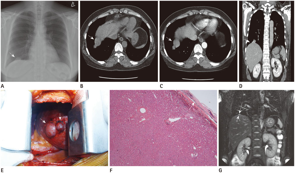
Fig. 1 A 37-year-old woman with intrathoracic ectopic liver. A. Chest radiograph showing a 3.5 cm sized mass (arrow) in the lower lobe of the right lung. The mass is ovoid in shape with smooth margins. B. Transverse unenhanced CT scan showing a lesion (arrow) with a density of 54 ± 4.3 Hounsfield units, similar to liver tissue. C. Chest CT with contrast enhancement showing mass with smooth margins (arrow) abutting the diaphragm and liver. The mass and liver are isodense during the arterial phase. D. Coronal reconstructed chest CT with contrast enhancement during the delayed phase showing a subpleural mass (arrow) abutting the diaphragm with isodense enhancement to liver tissue. E. Intraoperative view showing the 'mushroom-like' appearance of the intrathoracic ectopic liver (arrows) over the diaphragm. F. Histological examination, showing that the lesion is relatively well encapsulated (arrow), with histologic features similar to those of normal hepatic parenchyma (hematoxylin and eosin stain, × 40). G. One year follow-up coronal MRI with half-Fourier acquisition single-shot turbo spin-echo showing a small amount of focal fluid collection of high signal intensity (long arrow) at the site of previous masses on a hypointense thin band of the intact diaphragm (short arrow).
Reference
-
1. Le Roux BT. Heterotopic intrathoracic liver. Thorax. 1961; 16:68–71.2. Chen F, Heller DS, Bethel C, Faye-Petersen O. Intrathoracic ectopic lobe of liver presenting as pulmonary sequestration. Fetal Pediatr Pathol. 2005; 24:155–159.3. Iber T, Rintala R. Intrapulmonary ectopic liver. J Pediatr Surg. 1999; 34:1425–1426.4. Bedii Salman A. Left-sided congenital diaphragmatic hernia associated with intrathoracic ectopic liver lobule. Eur J Cardiothorac Surg. 2002; 21:558–560.5. Dinkel HP, Lorenz MH, Stein R, Kolb M. Transdiaphragmatic liver herniation mimicking pulmonary nodule. Eur J Radiol Extra. 2003; 46:17–20.6. An J, Han J, Lee KS, Choi YS. Supradiaphragmatic heterotopic liver presenting as a pleural mass: a case report. Tuberc Respir Dis. 2010; 69:191–195.7. Leone N, De Paolis P, Carrera M, Carucci P, Musso A, David E, et al. Ectopic liver and hepatocarcinogenesis: report of three cases with four years' follow-up. Eur J Gastroenterol Hepatol. 2004; 16:731–735.8. Collan Y, Hakkiluoto A, Hästbacka J. Ectopic liver. Ann Chir Gynaecol. 1978; 67:27–29.9. Mendoza A, Voland J, Wolf P, Benirschke K. Supradiaphragmatic liver in the lung. Arch Pathol Lab Med. 1986; 110:1085–1086.10. Iochum S, Ludig T, Walter F, Sebbag H, Grosdidier G, Blum AG. Imaging of diaphragmatic injury: a diagnostic challenge? Radiographics. 2002; 22 Spec No:S103–S116. discussion S116-S118.
- Full Text Links
-
- Actions
-
Cited
- CITED
-
- Close
- Share
-
- Similar articles
-
- A case of intrathoracic ectopic kidney presenting with congenital diaphragmatic hernia
- Intrathoracic Ectopic Live: A case report
- Impaction of an intrathoracic kidney acted as a shield against herniation of the abdominal viscera in a case of right congenital diaphragmatic hernia
- Supradiaphragmatic Heterotopic Liver Presenting as a Pleural Mass: A Case Report
- Congenital Thoracic Ectopic Kidney associated with Diaphragmatic Hernia in a 15-month-old Boy
