Chonnam Med J.
2016 May;52(2):123-127. 10.4068/cmj.2016.52.2.123.
Ureteral Stent Insertion in the Management of Renal Colic during Pregnancy
- Affiliations
-
- 1Department of Urology, CHA University Medical School, Seongnam, Korea. dsparkmd@cha.ac.kr
- KMID: 2164323
- DOI: http://doi.org/10.4068/cmj.2016.52.2.123
Abstract
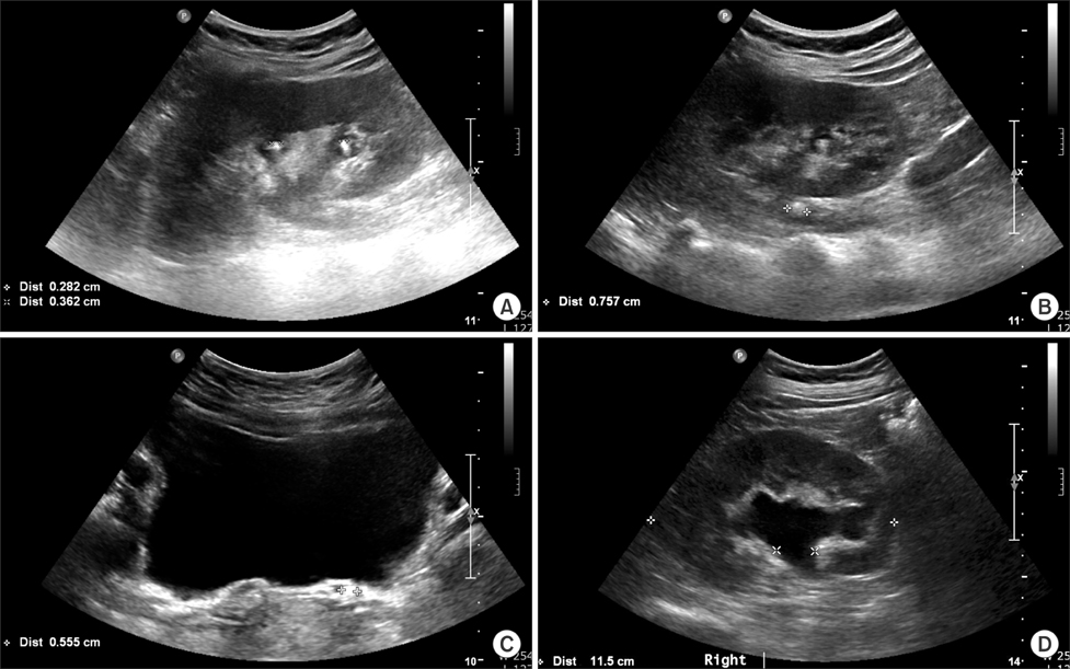
- To determine an optimal invasive intervention for renal colic patients during pregnancy after conservative treatments have been found to be unhelpful. Among the available invasive interventions, we investigated the reliability of a ureteral stent insertion, which is considered the least invasive intervention during pregnancy. Between June 2006 and February 2015, a total of 826 pregnant patients came to the emergency room or urology outpatient department, and 39 of these patients had renal colic. The mean patient age was 30.49 years. In this retrospective cohort study, the charts of the patients were reviewed to collect data that included age, symptoms, the lateralities and locations of urolithiasis, trimester, pain following treatment and pregnancy complications. Based on ultrasonography diagnoses, 13 patients had urolithiasis, and 13 patients had hydronephrosis without definite echogenicity of the ureteral calculi. Conservative treatments were successful in 25 patients. Among these treatments, antibiotics were used in 15 patients, and the remaining patients received only hydration and analgesics without antibiotics. Several urological interventions were required in 14 patients. The most common intervention was ureteral stent insertion, which was performed in 13 patients to treat hydronephrosis or urolithiasis. The patients' pain was relieved following these interventions. Only one patient received percutaneous nephrostomy due to pyonephrosis. No pregnancy complications were noted. Ureteral stent insertion is regarded as a reliable and stable first-line urological intervention for pregnant patients with renal colic following conservative treatments. Ureteral stent insertion has been found to be equally effective and safe as percutaneous nephrostomy, which is associated with complications that include bleeding and dislocation, and the inconvenience of using external drainage system.
Keyword
MeSH Terms
-
Analgesics
Anti-Bacterial Agents
Cohort Studies
Diagnosis
Dislocations
Drainage
Emergency Service, Hospital
Hemorrhage
Humans
Hydronephrosis
Nephrostomy, Percutaneous
Outpatients
Pregnancy Complications
Pregnancy*
Pyonephrosis
Renal Colic*
Retrospective Studies
Stents*
Ultrasonography
Ureter*
Ureteral Calculi
Urinary Catheters
Urolithiasis
Urology
Analgesics
Anti-Bacterial Agents
Figure
Reference
-
1. Andreoiu M, MacMahon R. Renal colic in pregnancy: lithiasis or physiological hydronephrosis? Urology. 2009; 74:757–761.
Article2. Srirangam SJ, Hickerton B, Van Cleynenbreugel B. Management of urinary calculi in pregnancy: a review. J Endourol. 2008; 22:867–875.
Article3. Wayment RO, Schwartz BF. Pregnancy and Urolithiasis [Internet]. USA: Emedicine;c2009. Updated 2015 Apr 17. Cited 2014 Nov 19. Available from: http://emedicine.medscape.com/article/455830-overview/.4. Gorton E, Whitfield HN. Renal calculi in pregnancy. Br J Urol. 1997; 80:Suppl 1. 4–9.5. Biyani CS, Joyce AD. Urolithiasis in pregnancy. II: management. BJU Int. 2002; 89:819–823.
Article6. Charalambous S, Fotas A, Rizk DE. Urolithiasis in pregnancy. Int Urogynecol J Pelvic Floor Dysfunct. 2009; 20:1133–1136.
Article7. Peake SL, Roxburgh HB, Langlois SL. Ultrasonic assessment of hydronephrosis of pregnancy. Radiology. 1983; 146:167–170.
Article8. Roberts JA. Hydronephrosis of pregnancy. Urology. 1976; 8:1–4.
Article9. Hendricks SK, Ross SO, Krieger JN. An algorithm for diagnosis and therapy of management and complications of urolithiasis during pregnancy. Surg Gynecol Obstet. 1991; 172:49–54.
Article10. Juan YS, Wu WJ, Chuang SM, Wang CJ, Shen JT, Long CY, et al. Management of symptomatic urolithiasis during pregnancy. Kaohsiung J Med Sci. 2007; 23:241–246.
Article11. Butler EL, Cox SM, Eberts EG, Cunningham FG. Symptomatic nephrolithiasis complicating pregnancy. Obstet Gynecol. 2000; 96:753–756.
Article12. Patel SJ, Reede DL, Katz DS, Subramaniam R, Amorosa JK. Imaging the pregnant patient for nonobstetric conditions: algorithms and radiation dose considerations. Radiographics. 2007; 27:1705–1722.
Article13. Swanson SK, Heilman RL, Eversman WG. Urinary tract stones in pregnancy. Surg Clin North Am. 1995; 75:123–142.
Article14. Dhar M, Denstedt JD. Imaging in diagnosis, treatment, and follow-up of stone patients. Adv Chronic Kidney Dis. 2009; 16:39–47.
Article15. Evans HJ, Wollin TA. The management of urinary calculi in pregnancy. Curr Opin Urol. 2001; 11:379–384.
Article16. Scarpa RM, De Lisa A, Usai E. Diagnosis and treatment of ureteral calculi during pregnancy with rigid ureteroscopes. J Urol. 1996; 155:875–877.
Article17. Travassos M, Amselem I, Filho NS, Miguel M, Sakai A, Consolmagno H, et al. Ureteroscopy in pregnant women for ureteral stone. J Endourol. 2009; 23:405–407.
Article18. Johnson EB, Krambeck AE, White WM, Hyams E, Beddies J, Marien T, et al. Obstetric complications of ureteroscopy during pregnancy. J Urol. 2012; 188:151–154.
Article19. Lifshitz DA, Lingeman JE. Ureteroscopy as a first-line intervention for ureteral calculi in pregnancy. J Endourol. 2002; 16:19–22.
Article20. Shah A, Chandak P, Tiptaft R, Glass J, Dasgupta P. Percutaneous nephrolithotomy in early pregnancy. Int J Clin Pract. 2004; 58:809–810.
Article21. Tóth C, Tóth G, Varga A, Flaskó T, Salah MA. Percutaneous nephrolithotomy in early pregnancy. Int Urol Nephrol. 2005; 37:1–3.
Article22. Streem SB. Contemporary clinical practice of shock wave lithotripsy: a reevaluation of contraindications. J Urol. 1997; 157:1197–1203.
Article


