Korean J Perinatol.
2015 Dec;26(4):373-376. 10.14734/kjp.2015.26.4.373.
Spontaneous Uterine Rupture during Second Trimester Pregnancy after High-intensity Focused Ultrasound
- Affiliations
-
- 1Department of Obstetrics and Gynecology, Dankook University College of Medicine, Cheonan, Korea. pch10@dankook.ac.kr
- KMID: 2164197
- DOI: http://doi.org/10.14734/kjp.2015.26.4.373
Abstract
- Uterine rupture during pregnancy does not occur frequently, but is associated with high rates of maternal and perinatal morbidity and mortality. As a non-invasive and conservative approach, high-intensity focused ultrasound (HIFU) has received attention from both gynecologists and patients for the treatment of fibroids, especially women who wish to preserve uterus. However, there are not enough studies about complication and prognosis related pregnancy after HIFU. We present a case of uterine rupture that occurred in second trimester who had been HIFU 3months before pregnancy.
MeSH Terms
Figure
-
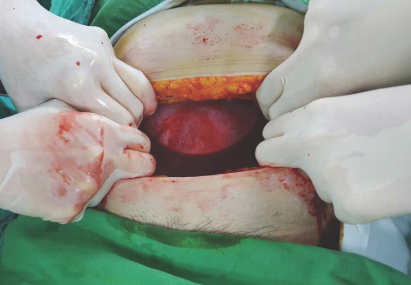
Fig. 1. Hematoma was exposed directly after entering abdominal cavity.
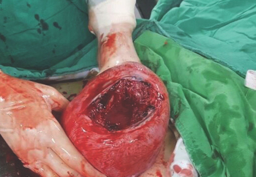
Fig. 2. The uterus was ruptured and split up about 10 cm at anterior fundus.
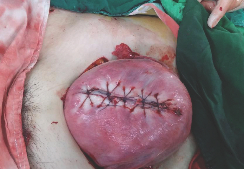
Fig. 3. Operative findings showing repaired uterus.
Cited by 2 articles
-
Effects of a video education program for patients with benign uterine tumors receiving high-intensity focused ultrasound treatment
Mi Suk Hong, Hyoung Sook Park, Young Suk Cho
Korean J Women Health Nurs. 2020;26(2):151-160. doi: 10.4069/kjwhn.2020.03.31.The Effects of Myoma and Uterine Preserving Procedures for Myoma on Pregnancy Outcomes
Gi Soo Um, Hyun Sun Ko
J Korean Soc Matern Child Health. 2022;26(2):45-51. doi: 10.21896/jksmch.2022.26.2.45.
Reference
-
References
1. Kennedy JE. High-intensity focused ultrasound in the treatment of solid tumours. Nat Rev Cancer. 2005; 5:321–7.
Article2. Hahn ST. High-Intensity focused ultrasound in the solid tumor treatment. J Korean Med Assoc. 2006; 49:707–16.
Article3. Fry WJ, Mosberg WH Jr, Barnard JW, Fry FJ. Production of focal destructive lesions in the central nervous system with ultrasound. J Neurosurg. 1954; 11:471–8.
Article4. Stewart EA, Gedroyc WM, Tempany CM, Quade BJ, Inbar Y, Ehrenstein T, et al. Focused ultrasound treatment of uterine fibroid tumors: safety and feasibility of a noninvasive thermoablative technique. Am J Obstet Gynecol. 2003; 189:48–54.
Article5. Tempany CM, Stewart EA, McDannold N, Quade BJ, Jolesz FA, Hynynen K. MR imaging-guided focused ultrasound surgery of uterine leiomyomas: a feasibility study. Radiology. 2003; 226:897–905.
Article6. Hindley J, Gedroyc WM, Regan L, Stewart E, Tempany C, Hynyen K, et al. MRI guidance of focused ultrasound therapy of uterine fibroids: early results. AJR Am J Roentgenol. 2004; 183:1713–9.7. Fennessy FM, Tempany CM, McDannold NJ, So MJ, Hesley G, Gostout B, et al. Uterine leiomyomas: MR imaging-guided focused ultrasound surgery–results of different treatment protocols. Radiology. 2007; 243:885–93.8. Qin J, Chen JY, Zhao WP, Hu L, Chen WZ, Wang ZB. Outcome of unintended pregnancy after ultrasound-guided high-intensity focused ultrasound ablation of uterine fibroids. Int J Gynaecol Obstet. 2012; 117:273–7.
Article9. Bohlmann MK, Hoellen F, Hunold P, David M. High-intensity focused ultrasound ablation of uterine fibroids – potential impact on fertility and pregnancy outcome. Geburtshilfe Frauenheilkd. 2014; 74:139–45.
Article10. Li XW, Liang MY, Wang JL, Wang DP. Spontaneous uterine rupture during late pregnancy after high-intensity focused ultrasound. Chin Med J. 2015; 128:1419.
Article
- Full Text Links
-
- Actions
-
Cited
- CITED
-
- Close
- Share
-
- Similar articles
-
- Repetitive Spontaneous Uterine Rupture in the First Trimester after Laparoscopic Myomectomy: A Case Report and Review of Literature
- A Case of Spontaneous Uterine Rupture in the Second Trimester of Pregnancy
- A Case of Uterine Rupture in the 18th Week of Pregnancy
- A Case of Spontaneous Uterine Rupture of the Unscarred Uterus in 14 Weeks Gestation
- Spontaneous uterine rupture during second trimester
