Acetabular Reconstruction in Total Hip Arthroplasty
- Affiliations
-
- 1Department of Orthopedic Surgery, Korea University Guro Hospital, Seoul, Korea. shonwy@hotmail.com
- KMID: 2161309
- DOI: http://doi.org/10.5371/hp.2016.28.1.1
Abstract
- The difficulties encountered in dealing with the bone deficient acetabulum are amongst the greatest challenges in hip surgery. Acetabular reconstruction in revision total hip arthroplasty can successfully be achieved with hemispherical components featuring a porous or roughened ingrowth surface and options for placement of multiple screws for minor acetabular defect. Acetabular component selection is mostly based on the amount of bone loss present. In the presence of combined cavitary and segmental defects without superior acetabular coverage, reconstructions with a structural acetabular allograft protected by a cage or a custom-made triflange cage have been one of preferred surgical options. The use of a cage or ring over structural allograft bone for massive uncontained defects in acetabular revision can restore host bone stock and facilitate subsequent rerevision surgery to a certain extent. But high complication rates have been reported including aseptic loosening, infection, dislocation and metal failure. On the other hand, recent literature is reporting satisfactory outcomes with the use of modular augments combined with a hemispherical shell for major acetabular defect. Highly porous metals have been introduced for clinical use in arthroplasty surgery over the last decade. Their higher porosity and surface friction are ideal for acetabular revision, optimizing biological fixation. The use of trabecular metal cups in acetabular revision has yielded excellent clinical results. This article summarizes author's experience regarding revision acetabular reconstruction options following failed hip surgery including arthroplasty.
MeSH Terms
Figure
-
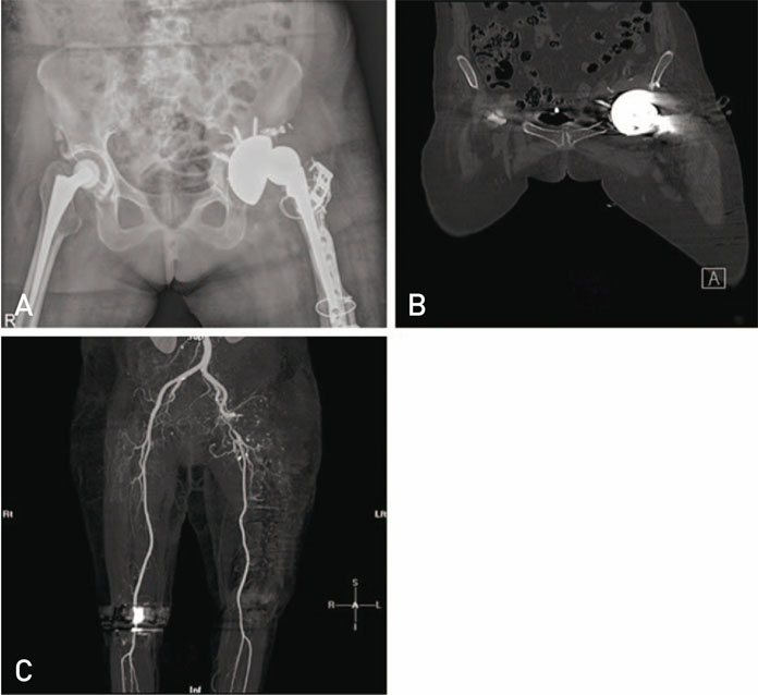
Fig. 1 (A, B) Radiograph and computed tomography (CT) scan respectively showing intra-pelvic acetabular protrusion with screw. (C) This patient sustained injury to the external iliac artery during acetabular component retrieval which is seen in the CT angiogram image.
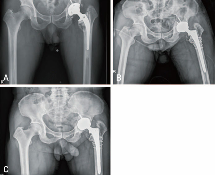
Fig. 2 (A) Radiograph showing lysis around acetabular and femoral component. (B) Radiograph taken after revision with cementless acetabular and femoral components. (C) Follow up radiograph at 2 years showing loosening of acetabular component.
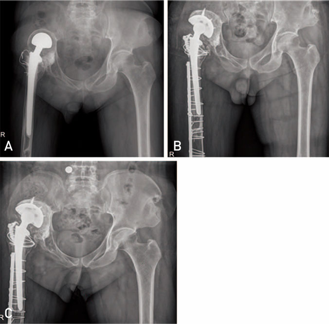
Fig. 3 (A) Radiograph showing severe cavitary defect in the right acetabulam. (B) Follow up radiograph at one year after revision done using impaction bone grafting and cementless acetabular cup. (C) Follow up radiograph at 3 years showing superior migration of cementless acetabular cup with resorption of the allograft.
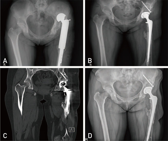
Fig. 4 (A) Radiograph showing a failed bipolar tumor prosthesis with a severe superior acetabular defect. (B) Radiograph taken after revision with structural allograft and a cementless acetabular cup. (C) Computed tomography scan image showing well incorporated graft, 6 years after revision surgery. (D) Radiograph taken at 8 years after revision surgery showing well-functioning implants with no evidence of loosening.
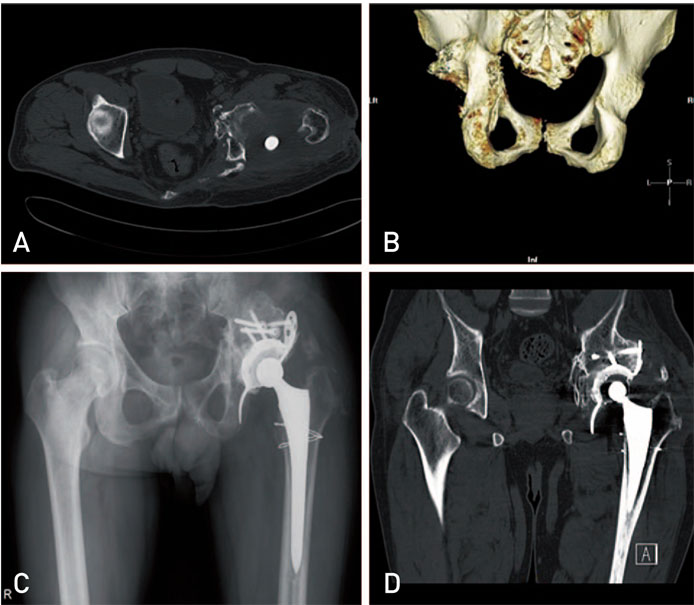
Fig. 5 (A, B) Computed tomography (CT) scan images showing severe bone defect in the postero-superior column of acetabulam. (C) Radiograph taken after revision with structural allograft and anti-protrusio cage. (D) CT scan image done after 1 year showing a good fixation of cage between host bone and structural allograft.
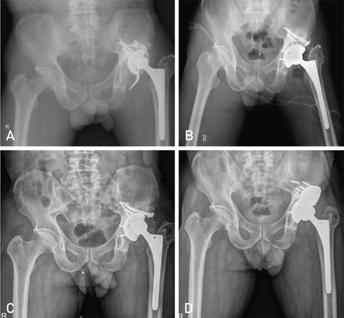
Fig. 6 (A) Radiograph showing lysis and loosening of the anti-protrusio cage. (B) Post-operative radiograph done after revision with structural allograft and a cementless acetabular cup. (C) Follow up radiograph at 4 years, showing loosening of the acetabular cup. (D) Post-operative radiograph taken after revision with tantalum metal block and cementless acetabular cup.
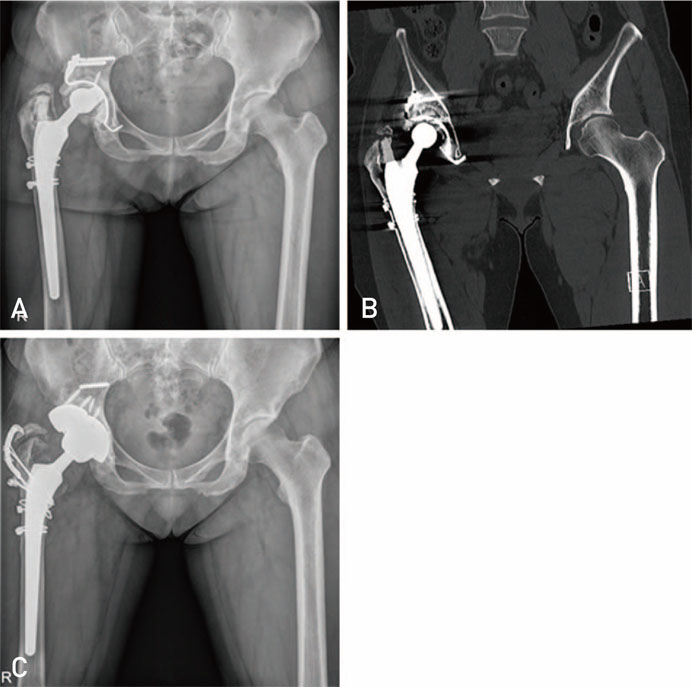
Fig. 7 (A, B) Radiograph and computed tomography respectively showing failure of Kerboull plate because of fracture of screw at 11 years after operation. (C) Post-operative radiograph after revision with metal augment.
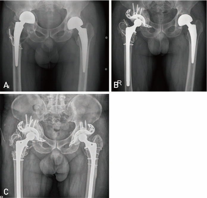
Fig. 8 (A) Radiograph showing superior and medial migration of bipolar prosthesis. (B) Radiograph taken after revision with impaction bone grafting and anti-protrusio cage. (C) Follow up radiograph at 10 years showing a well organised graft with no evidence of implant loosening.
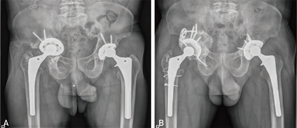
Fig. 9 (A) Radiograph showing supero-medial migration of acetabular component. (B) Follow up radiograph at 1 year postoperaion showing no resorption of allograft and a good functioning anti-protrusio cage.
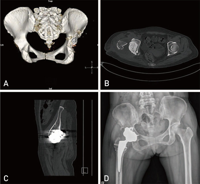
Fig. 10 (A, B, C) Computed tomography scan images showing postero-superior column defect in the acetabulam. (D) Postoperative radiograph done after revision with tantalum metal block and a cementless acetabular cup.
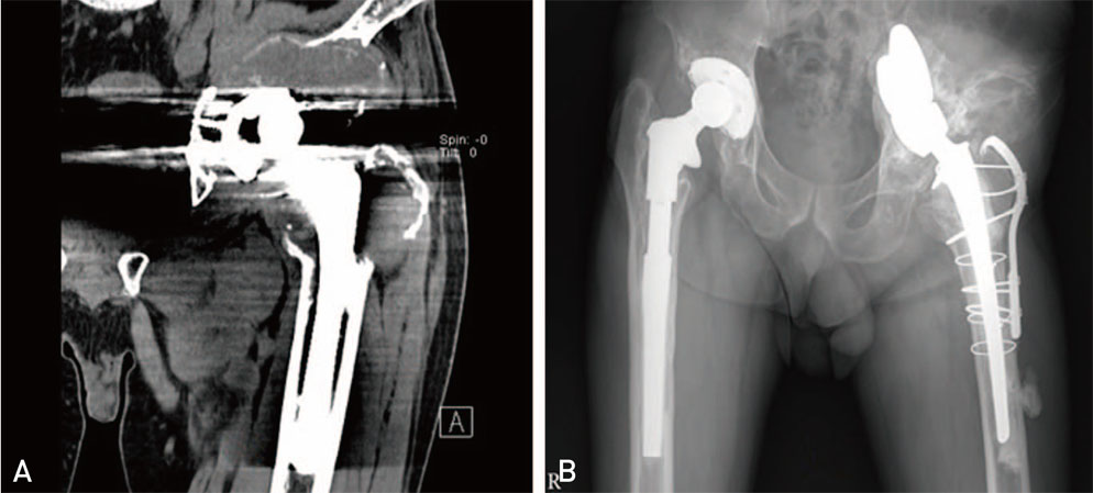
Fig. 11 (A) Computed tomography scan image showing pelvic discontinuity due to severe osteolysis. (B) Follow up radiograph at 6 years after revision with saddle prosthesis.
Reference
-
1. Slooff TJ, Schimmel JW, Buma P. Cemented fixation with bone grafts. Orthop Clin North Am. 1993; 24:667–677.2. van Egmond N, De Kam DC, Gardeniers JW, Schreurs BW. Revisions of extensive acetabular defects with impaction grafting and a cement cup. Clin Orthop Relat Res. 2011; 469:562–573.3. van Haaren EH, Heyligers IC, Alexander FG, Wuisman PI. High rate of failure of impaction grafting in large acetabular defects. J Bone Joint Surg Br. 2007; 89:296–300.
Article4. Bostrom MP, Lehman AP, Buly RL, Lyman S, Nestor BJ. Acetabular revision with the Contour antiprotrusio cage: 2- to 5-year followup. Clin Orthop Relat Res. 2006; 453:188–194.
Article5. Issack PS, Nousiainen M, Beksac B, Helfet DL, Sculco TP, Buly RL. Acetabular component revision in total hip of major bone loss and pelvic discontinuity. Am J Orthop (Belle Mead NJ). 2009; 38:550–556.6. Sporer SM. How to do a revision total hip arthroplasty: revision of the acetabulum. J Bone Joint Surg Am. 2011; 93:1359–1366.7. Borland WS, Bhattacharya R, Holland JP, Brewster NT. Use of porous trabecular metal augments with impaction bone grafting in management of acetabular bone loss. Acta Orthop. 2012; 83:347–352.
Article8. Abolghasemian M, Tangsataporn S, Sternheim A, Backstein D, Safir O, Gross AE. Combined trabecular metal acetabular shell and augment for acetabular revision with substantial bone loss: a mid-term review. Bone Joint J. 2013; 95:166–172.
Article9. Yun HH, Shon WY, Hong SJ, Yoon JR, Yang JH. Relationship between the pelvic osteolytic volume on computed tomography and clinical outcome in patients with cementless acetabular components. Int Orthop. 2011; 35:1453–1459.
Article10. Suh DH, Han SB, Yun HH, Chun SK, Shon WY. Characterization of progression of pelvic osteolysis after cementless total hip arthroplasty: computed tomographic study. J Arthroplasty. 2013; 28:1851–1855.
Article11. Reiley MA, Bond D, Branick RI, Wilson EH. Vascular complications following total hip arthroplasty. A review of the literature and a report of two cases. Clin Orthop Relat Res. 1984; (186):23–28.
Article12. Patel S, Sukeik M, Haddad FS. Initial implant stability predicts migration but not failure in cementless acetabular revision with bone grafting. J Arthroplasty. 2013; 28:832–837.
Article13. Mazhar Tokgözoğlu A, Aydin M, Atilla B, Caner B. Scintigraphic evaluation of impaction grafting for total hip arthroplasty revision. Arch Orthop Trauma Surg. 2000; 120:416–419.
Article14. Goldberg VM. Selection of bone grafts for revision total hip arthroplasty. Clin Orthop Relat Res. 2000; (381):68–76.
Article15. Stevenson S. Biology of bone grafts. Orthop Clin North Am. 1999; 30:543–552.
Article16. Schimmel JW, Buma P, Versleyen D, Huiskes R, Slooff TJ. Acetabular reconstruction with impacted morselized cancellous allografts in cemented hip arthroplasty: a histological and biomechanical study on the goat. J Arthroplasty. 1998; 13:438–448.
Article17. Etienne G, Ragland PS, Mont MA. Use of cancellous bone chips and demineralized bone matrix in the treatment of acetabular osteolysis: preliminary 2-year follow-up. Orthopedics. 2004; 27:1 Suppl. s123–s126.
Article18. Jasty M, Harris WH. Total hip reconstruction using frozen femoral head allografts in patients with acetabular bone loss. Orthop Clin North Am. 1987; 18:291–299.
Article19. Avci S, Connors N, Petty W. 2- to 10-year follow-up study of acetabular revisions using allograft bone to repair bone defects. J Arthroplasty. 1998; 13:61–69.
Article20. Buttaro MA, Comba F, Pusso R, Piccaluga F. Acetabular revision with metal mesh, impaction bone grafting, and a cemented cup. Clin Orthop Relat Res. 2008; 466:2482–2490.
Article21. Brubaker SM, Brown TE, Manaswi A, Mihalko WM, Cui Q, Saleh KJ. Treatment options and allograft use in revision total hip arthroplasty the acetabulum. J Arthroplasty. 2007; 22:7 Suppl 3. 52–56.22. Murphy SB. Management of acetabular bone stock deficiency. J Arthroplasty. 2005; 20:4 Suppl 2. 85–90.23. Paprosky WG, Magnus RE. Principles of bone grafting in revision total hip arthroplasty. Acetabular technique. Clin Orthop Relat Res. 1994; (298):147–155.
Article24. Sporer SM, O'Rourke M, Chong P, Paprosky WG. The use of structural distal femoral allografts for acetabular reconstruction. Average ten-year follow-up. J Bone Joint Surg Am. 2005; 87:760–765.
Article25. O'Rourke MR, Paprosky WG, Rosenberg AG. Use of structural allografts in acetabular revision surgery. Clin Orthop Relat Res. 2004; (420):113–121.
Article26. Pollock FH, Whiteside LA. The fate of massive allografts in total hip acetabular revision surgery. J Arthroplasty. 1992; 7:271–276.
Article27. Garbuz D, Morsi E, Gross AE. Revision of the acetabular component of a total hip arthroplasty with a massive structural allograft. Study with a minimum five-year follow-up. J Bone Joint Surg Am. 1996; 78:693–697.
Article28. Gross AE. Restoration of acetabular bone loss 2005. J Arthroplasty. 2006; 21:4 Suppl 1. 117–120.
Article29. Gross AE, Goodman S. The current role of structural grafts and cages in revision arthroplasty of the hip. Clin Orthop Relat Res. 2004; (429):193–200.30. Oakes DA, Cabanela ME. Impaction bone grafting for revision hip arthroplasty: biology and clinical applications. J Am Acad Orthop Surg. 2006; 14:620–628.31. Patil N, Hwang K, Goodman SB. Cancellous impaction bone grafting of acetabular defects in complex primary and revision total hip arthroplasty. Orthopedics. 2012; 35:e306–e312.
Article32. Zehntner MK, Ganz R. Midterm results (5.5-10 years) of acetabular allograft reconstruction with the acetabular reinforcement ring during total hip revision. J Arthroplasty. 1994; 9:469–479.
Article33. Tägil M. The morselized and impacted bone graft. Animal experiments on proteins, impaction and load. Acta Orthop Scand Suppl. 2000; 290:1–40.
Article34. Jasty M, Harris WH. Salvage total hip reconstruction in patients with major acetabular bone deficiency using structural femoral head allografts. J Bone Joint Surg Br. 1990; 72:63–67.
Article35. Berry DJ, Müller ME. Revision arthroplasty using an antiprotrusio cage for massive acetabular bone deficiency. J Bone Joint Surg Br. 1992; 74:711–715.
Article36. Peters CL, Curtain M, Samuelson KM. Acetabular revision with the Burch-Schnieder antiprotrusio cage and cancellous allograft bone. J Arthroplasty. 1995; 10:307–312.
Article37. Gill TJ, Sledge JB, Müller ME. The Bürch-Schneider antiprotrusio cage in revision total hip arthroplasty: indications, principles and long-term results. J Bone Joint Surg Br. 1998; 80:946–953.
Article38. Regis D, Sandri A, Bonetti I, Bortolami O, Bartolozzi P. A minimum of 10-year follow-up of the Burch-Schneider cage and bulk allografts for the revision of pelvic discontinuity. J Arthroplasty. 2012; 27:1057–1063.e1.
Article39. Nehme A, Lewallen DG, Hanssen AD. Modular porous metal augments for treatment of severe acetabular bone loss during revision hip arthroplasty. Clin Orthop Relat Res. 2004; (429):201–208.
Article40. Bobyn JD, Stackpool GJ, Hacking SA, Tanzer M, Krygier JJ. Characteristics of bone ingrowth and interface mechanics of a new porous tantalum biomaterial. J Bone Joint Surg Br. 1999; 81:907–914.
Article41. Van Kleunen JP, Lee GC, Lementowski PW, Nelson CL, Garino JP. Acetabular revisions using trabecular metal cups and augments. J Arthroplasty. 2009; 24:6 Suppl. 64–68.
Article42. Ballester Alfaro JJ, Sueiro Fernández J. Trabecular Metal buttress augment and the Trabecular Metal cup-cage construct in revision hip arthroplasty for severe acetabular bone loss and pelvic discontinuity. Hip Int. 2010; 20:Suppl 7. S119–S127.
Article43. Malhotra R, Kancherla R, Kumar V, Soral A. Trabecular metal acetabular revision system (cup-cage construct) to address the massive acetabular defects in revision arthroplasty. Indian J Orthop. 2012; 46:483–486.
Article44. Jansen JA, van de Sande MA, Dijkstra PD. Poor long-term clinical results of saddle prosthesis after resection of periacetabular tumors. Clin Orthop Relat Res. 2013; 471:324–331.
Article
