J Korean Soc Radiol.
2015 Mar;72(3):164-170. 10.3348/jksr.2015.72.3.164.
The Value of MRI in Idiopathic Tarsal Tunnel Syndrome by Measuring the Cross-Sectional Area of Tarsal Tunnel
- Affiliations
-
- 1Department of Radiology, Gachon University, Gil Hospital, Incheon, Korea. youme34@gilhospital.com
- 2Department of Orthopedic Surgery, Gachon University, Gil Hospital, Incheon, Korea.
- KMID: 2161167
- DOI: http://doi.org/10.3348/jksr.2015.72.3.164
Abstract
- PURPOSE
The purpose of this study was to evaluate the use of MRI as a diagnostic test in tarsal tunnel syndrome. There are no published reports with this aim and no diagnostic standard for idiopathic tarsal tunnel syndrome (TTS) using imaging modalities.
MATERIALS AND METHODS
We retrospectively searched our Picture Archiving and Communication System data and medical records to identify patients who were clinically and electomyographically diagnosed with idiopathic TTS without space-occupying lesion on MRI. Twenty five patients were included in the patient group. Another twenty-five patients who underwent ankle MRI for Achilles tendon disease were selected and included in the control group. Cross-sectional areas (CSA) of tarsal tunnel were manually measured independently by two radiologists who were blinded to clinical and surgical results, using three-dimensional reconstruction software in our hospital. Measurements were done on axial images at three levels (level 1, tibiotalar joint level; level 2, medial malleolar tip level; level 3, sustentaculum tali level). Patient and control group data were statistically analyzed by the Mann-Whitney test.
RESULTS
The mean values of CSA at levels 1, 2, and 3 of the tarsal tunnel were 87.8 mm2, 98.2 mm2, and 105.2 mm2, respectively in the patient group; and 100.0 mm2, 113.8 mm2, and 127.9 mm2 in the control group, respectively, in reader 1; and 86.2 mm2, 97.6 mm2, 105.2 mm2, respectively in the patient group; and 99.7 mm2, 112.3 mm2, 124.4 mm2, respectively, in the control group, in reader 2. The mean CSA in the patient group was significantly less than that of the control group at all three levels (p < 0.05). Intra-class correlation coefficient value between reader 1 and reader 2 were 0.98 in group 1, and 0.97 in group 2, respectively.
CONCLUSION
MRI can be helpful in the assessment of idiopathic tarsal tunnel syndrome. CSA measurements of tarsal tunnel at each level may predict TTS even though there are no space occupying lesions in the tarsal tunnel on MRI.
MeSH Terms
Figure
-

Fig. 1 The three axial levels of the tarsal tunnel at which the cross-sectional area were measured. A. Cross section of ankle at level of tibiotalar joint. B. Cross section of ankle at level of medial malleolar tip. C. Cross section of ankle at level of sustentaculum tali.
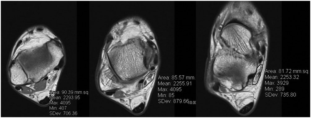
Fig. 2 MR axial image of 59/M with idiopathic tarsal tunnel syndrome, right ankle. Some of the patient group showed flattened appearance of fibro-osseous tunnel on the transverse images.
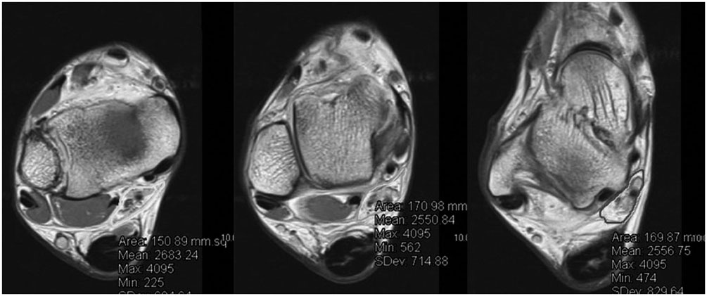
Fig. 3 MR axial image of 31/M. Right ankle MRI for postoperative follow-up 2 years after Achilles tendon repair.
Reference
-
1. Erickson SJ, Quinn SF, Kneeland JB, Smith JW, Johnson JE, Carrera GF, et al. MR imaging of the tarsal tunnel and related spaces: normal and abnormal findings with anatomic correlation. AJR Am J Roentgenol. 1990; 155:323–328.2. Narváez JA, Narváez J, Ortega R, Aguilera C, Sánchez A, Andía E. Painful heel: MR imaging findings. Radiographics. 2000; 20:333–352.3. Donovan A, Rosenberg ZS, Cavalcanti CF. MR imaging of entrapment neuropathies of the lower extremity. Part 2. The knee, leg, ankle, and foot. Radiographics. 2010; 30:1001–1019.4. Trepman E, Kadel NJ, Chisholm K, Razzano L. Effect of foot and ankle position on tarsal tunnel compartment pressure. Foot Ankle Int. 1999; 20:721–726.5. El Shazly O, El Shazly A, Desouky A, El Zohiery AK, Sakr HM. Anatomical bases of endoscopic tarsal tunnel release: anatomical and ultra-sonographic study with a preliminary clinical report. Surg Radiol Anat. 2011; 33:929–936.6. Kohno M, Takahashi H, Segawa H, Sano K. Neurovascular decompression for idiopathic tarsal tunnel syndrome: technical note. J Neurol Neurosurg Psychiatry. 2000; 69:87–90.7. Jung HJ, Lee SW, Jeong YM, Choi HY, Kim HS, Park HG, et al. The usefulness of the preoperative magnetic resonance imaging findings in the evaluation of tarsal tunnel syndrome. J Korean Soc Radiol. 2012; 66:183–192.8. Sora MC, Jilavu R, Grübl A, Genser-Strobl B, Staykov D, Seicean A. The posteromedial neurovascular bundle of the ankle: an anatomic study using plastinated cross sections. Arthroscopy. 2008; 24:258–263.e1.9. Martinoli C, Bianchi S, Gandolfo N, Valle M, Simonetti S, Derchi LE. US of nerve entrapments in osteofibrous tunnels of the upper and lower limbs. Radiographics. 2000; 20(Spec No):S199–S213. discussion S213-S217.10. Peer S, Kiechl G, Bonder G. Nerve compression syndromes. In : Peer S, Bodner G, editors. High-resolution sonography of the peripheral nervous system. Berlin: Springer;2003. p. 37.11. Cheung Y, Rosenberg ZS. MR imaging of the accessory muscles around the ankle. Magn Reson Imaging Clin N Am. 2001; 9:465–473. x.12. Kinoshita M, Okuda R, Morikawa J, Abe M. Tarsal tunnel syndrome associated with an accessory muscle. Foot Ankle Int. 2003; 24:132–136.13. Duncan I, Sullivan P, Lomas F. Sonography in the diagnosis of carpal tunnel syndrome. AJR Am J Roentgenol. 1999; 173:681–684.14. Klauser AS, Halpern EJ, De Zordo T, Feuchtner GM, Arora R, Gruber J, et al. Carpal tunnel syndrome assessment with US: value of additional cross-sectional area measurements of the median nerve in patients versus healthy volunteers. Radiology. 2009; 250:171–177.15. Dekel S, Papaioannou T, Rushworth G, Coates R. Idiopathic carpal tunnel syndrome caused by carpal stenosis. Br Med J. 1980; 280:1297–1299.16. Horch RE, Allmann KH, Laubenberger J, Langer M, Stark GB. Median nerve compression can be detected by magnetic resonance imaging of the carpal tunnel. Neurosurgery. 1997; 41:76–82. discussion 82-83.17. Bower JA, Stanisz GJ, Keir PJ. An MRI evaluation of carpal tunnel dimensions in healthy wrists: implications for carpal tunnel syndrome. Clin Biomech (Bristol, Avon). 2006; 21:816–825.18. Martins RS, Siqueira MG, Simplício H, Agapito D, Medeiros M. Magnetic resonance imaging of idiopathic carpal tunnel syndrome: correlation with clinical findings and electrophysiological investigation. Clin Neurol Neurosurg. 2008; 110:38–45.19. Ito T, Kijima M, Watanabe T, Sakuta M, Nishiyama K. Ultrasonography of the tibial nerve in vasculitic neuropathy. Muscle Nerve. 2007; 35:379–382.20. Lee D, Dauphinée DM. Morphological and functional changes in the diabetic peripheral nerve: using diagnostic ultrasound and neurosensory testing to select candidates for nerve decompression. J Am Podiatr Med Assoc. 2005; 95:433–437.21. Alshami AM, Cairns CW, Wylie BK, Souvlis T, Coppieters MW. Reliability and size of the measurement error when determining the cross-sectional area of the tibial nerve at the tarsal tunnel with ultrasonography. Ultrasound Med Biol. 2009; 35:1098–1102.22. Torres AL, Ferreira MC. Study of the anatomy of the tibial nerve and its branches in the distal medial leg. Acta Ortop Bras. 2012; 20:157–164.23. Ahn JH, Kim KJ, Kim HY, Choy WS, Yang DS. Surgical treatment of tarsal tunnel syndrome. J Korean Foot Ankle Soc. 2007; 11:187–191.
- Full Text Links
-
- Actions
-
Cited
- CITED
-
- Close
- Share
-
- Similar articles
-
- Tarsal Tunnel Syndrome associated with Os Sustentaculi (A Case Report)
- Treatment of Postoperative Tarsal Tunnel Syndrome with Autogeneous Vein Wrapping Graft
- Tarsal Tunnel Syndrome Associated with Gout Tophi: A Case Report
- Tarsal Tunnel Syndrome due to Varicose Veins Misdiagnosed as Ganglion Cyst: A Case Report
- Tarsal Tunnel Syndrome secondary to the Varicosis of Posterior Tibial Vein (Two Cases Report)
