Comparison of Distal Radius Fractures with or without Scaphoid Fractures
- Affiliations
-
- 1Department of Orthopedics, Yonsei University Wonju College of Medicine, Wonju, Korea. ojr1128@daum.net
- KMID: 2161160
- DOI: http://doi.org/10.12790/jkssh.2016.21.1.23
Abstract
- PURPOSE
Distal radius fracture is one of the most common factures, but incidence of concomitant scaphoid fracture is rare. The rarity makes diagnosing the concomitant scaphoid fracture often delayed. Thus, in this study, the authors examined the frequency of concomitant scaphoid injury in distal radius fracture and the type of distal radius fracture that is more commonly associated with simultaneous scaphoid fracture.
METHODS
We examined a total of 212 patients who had received treatment for the fracture in our institution. They were divided into two groups, isolated distal radius fracture group and distal radius fracture group with simultaneous scaphoid fracture, and their age, gender, body mass index and distal radius fracture type in accordance with AO classification were compared between the two groups.
RESULTS
Concomitant scaphoid fractures were found in 12 (5%) patients, and among them 10 cases were associated with type C distal radius fracture. Statistical comparison between the group with isolated distal radius fracture and the group with both distal radius and scaphoid fractures was made, and only comparison of distal radius fracture types showed statistical significance.
CONCLUSION
It is imperative to make timely and appropriate diagnosis of accompanying scaphoid fracture, since delay in making the diagnosis usually lead to many complications. We conclude that further diagnostic imaging such as computed tomography is necessary to make the correct diagnosis of concomitant scaphoid fracture, especially in type C distal radius fractures.
MeSH Terms
Figure
-
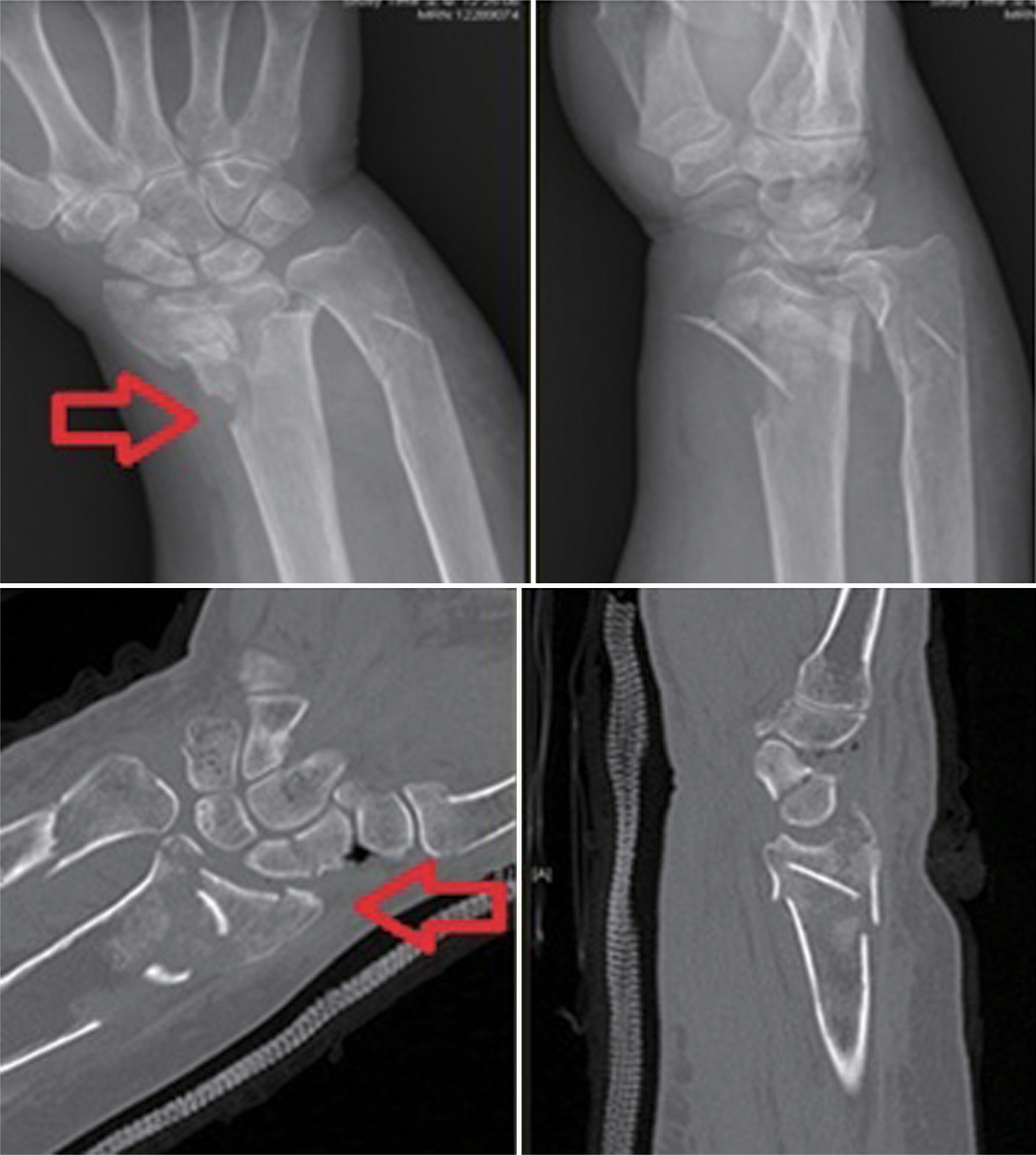
Fig. 1. This is a simple X-ray and computed tomography (CT) scan of a 69-year-old female patient admitted after a car accident. Displacement of scaphoid was not observed in the simple X-ray (1st arrow); however, fracture in both scaphoid and radio-ulnar bone was observed in the CT scan (2nd arrow).
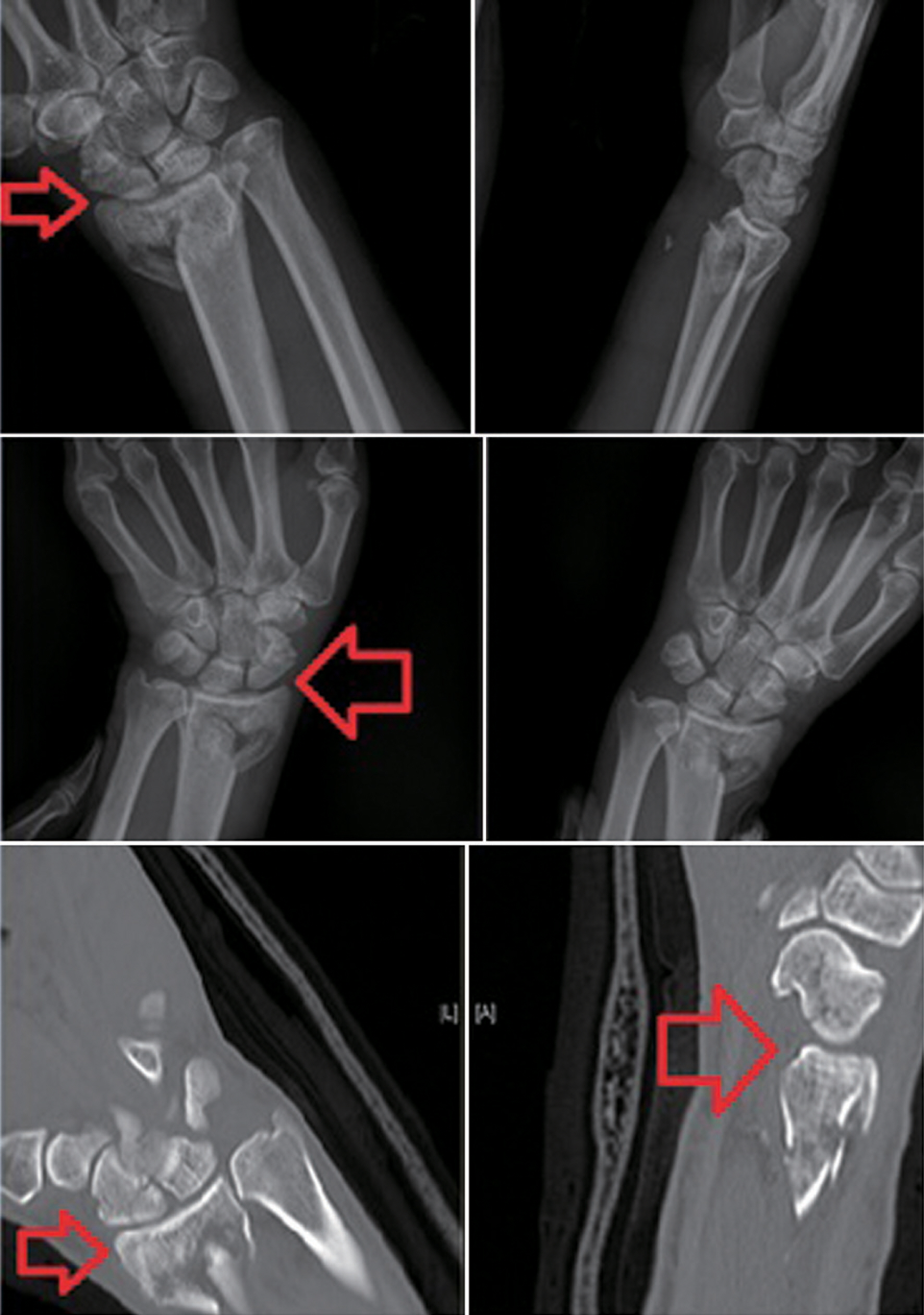
Fig. 2. This is a simple X-ray and computed tomography (CT) scan of a 51 -year-old male patient admitted after a falling down injury. After observing scaphoid displacement in simple X-ray (1st arrow), the wrist bending ulnar radius deviation was further examined. The scaphoid displacement became more distinctive with further examination (2nd arrow). Fracture in both scaphoid and distal radius was observed in the CT scan that followed (3rd, 4th arrow).
Reference
-
1. Wolfe SW. Distal radius fractures. Wolfe SW, Hotchkiss RN, Pederson WC, editors. Green's operative hand surgery. Philadelphia: Churchill Livingstone;2011. p. 561–638.
Article2. Chang CH, Tsai YS, Sun JS, Hou SM. Ipsilateral distal radius and scaphoid fractures. J Formos Med Assoc. 2000; 99:733–7.3. Hove LM. Simultaneous scaphoid and distal radial fractures. J Hand Surg Br. 1994; 19:384–8.
Article4. Moller BN. Simultaneous fracture of the carpal scaphoid and adjacent bones. Hand. 1983; 15:258–61.
Article5. Oskam J, De Graaf JS, Klasen HJ. Fractures of the distal radius and scaphoid. J Hand Surg Br. 1996; 21:772–4.
Article6. Pretell-Mazzini J, Carrigan RB. Simultaneous distal radial fractures and carpal bones injuries in children: a review article. J Pediatr Orthop B. 2011; 20:330–3.7. Downing ND, Karantana A. A revolution in the management of fractures of the distal radius. J Bone Joint Surg Br. 2008; 90:1271–5.
Article8. Kwon BC, Baek GH. Fluoroscopic diagnosis of scapholunate interosseous ligament injuries in distal radius fractures. Clin Orthop Relat Res. 2008; 466:969–76.
Article9. Sammer DM, Shah HM, Shauver MJ, Chung KC. The effect of ulnar styloid fractures on patient-rated outcomes after volar locking plating of distal radius fractures. J Hand Surg Am. 2009; 34:1595–602.
Article10. Smith JT, Keeve JP, Bertin KC, Mann RJ. Simultaneous fractures of the distal radius and scaphoid. J Trauma. 1988; 28:676–9.
Article11. Vukov V, Ristic K, Stevanovic M, Bumbasirevic M. Simultaneous fractures of the distal end of the radius and the scaphoid bone. J Orthop Trauma. 1988; 2:120–3.
Article12. Vigler M, Aviles A, Lee SK. Carpal fractures excluding the scaphoid. Hand Clin. 2006; 22:501–16.
Article13. Welling RD, Jacobson JA, Jamadar DA, Chong S, Caoili EM, Jebson PJ. MDCT and radiography of wrist fractures: radiographic sensitivity and fracture patterns. AJR Am J Roentgenol. 2008; 190:10–6.
Article14. Kiuru MJ, Haapamaki VV, Koivikko MP, Koskinen SK. Wrist injuries; diagnosis with multidetector CT. Emerg Radiol. 2004; 10:182–5.
Article15. Heo YM, Kim SB, Yi JW, et al. Evaluation of associated carpal bone fractures in distal radial fractures. Clin Orthop Surg. 2013; 5:98–104.
Article16. Cho CH, Son ES. Concomitant carpal injuries in distal radius fractures: restrospective analysis by plain radiographs and computed tomography. J Korean Fract Soc. 2015; 28:1–7.
- Full Text Links
-
- Actions
-
Cited
- CITED
-
- Close
- Share
-
- Similar articles
-
- Pediatric Fractures around the Wrist
- Ipsilateral Distal Radius and Scaphoid Fractures Associated with Posteromedial Dislocation of the Elbow Joint: A Case Report
- Operative Treatment of Carpal Scaphoid Fractures with Herbert Screw
- Complications of Distal Radius Fracture
- Arthroscopically Assisted Reduction of Distal Radius Fractures
