Cone-beam CT analysis of patients with obstructive sleep apnea compared to normal controls
- Affiliations
-
- 1Division of Radiology, Department of Oral Health and Diagnostic Sciences, Dental College of Georgia, Augusta University, Augusta, GA, USA. ahunter@gru.edu
- 2Park Avenue Oral and Facial Surgery, New York, NY, USA.
- 3Department of Biostatistics and Epidemiology, Augusta University Medical College of Georgia, Augusta, GA, USA.
- 4Department of Oral Health and Diagnostic Sciences, Georgia Regents University, Dental College of Georgia, Augusta, GA, USA.
- 5Division of Oral Medicine, Department of Oral Health and Diagnostic Sciences, Dental College of Georgia, Augusta University, Augusta, GA, USA.
- KMID: 2160152
- DOI: http://doi.org/10.5624/isd.2016.46.1.9
Abstract
- PURPOSE
To evaluate the upper airway dimensions of obstructive sleep apnea (OSA) and control subjects using a cone-beam computed tomography (CBCT) unit commonly applied in clinical practice in order to assess airway dimensions in the same fashion as that routinely employed in a clinical setting.
MATERIALS AND METHODS
This was a retrospective analysis utilizing existing CBCT scans to evaluate the dimensions of the upper airway in OSA and control subjects. The CBCT data of sixteen OSA and sixteen control subjects were compared. The average area, average volume, total volume, and total length of the upper airway were computed. Width and anterior-posterior (AP) measurements were obtained on the smallest axial slice.
RESULTS
OSA subjects had a significantly smaller average airway area, average airway volume, total airway volume, and mean airway width. OSA subjects had a significantly larger airway length measurement. The mean A-P distance was not significantly different between groups.
CONCLUSION
OSA subjects have a smaller upper airway compared to controls with the exception of airway length. The lack of a significant difference in the mean A-P distance may indicate that patient position during imaging (upright vs. supine) can affect this measurement. Comparison of this study with a future prospective study design will allow for validation of these results.
MeSH Terms
Figure
-
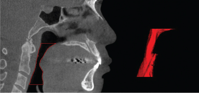
Fig. 1 Red area indicates the segmented airway with three-dimensional reconstruction.

Fig. 2 Linear measurements acquired on an obstructive sleep apnea subject. A. Sagittal view denoting the location of the smallest cross-sectional slice. B. Anterior-posterior distance, measured on the axial image. C. Width (lateral), measured on the axial image.
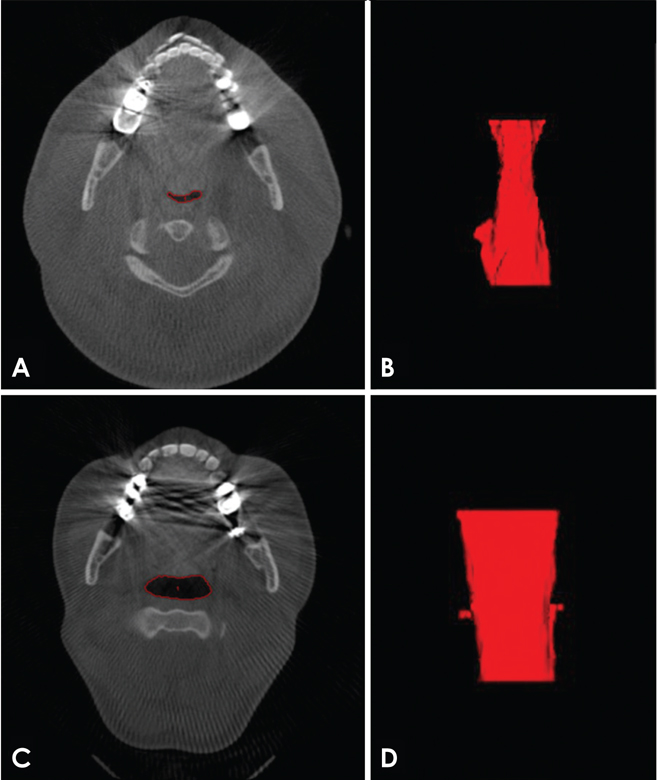
Fig. 3 Comparison of airway width (lateral) between an obstructive sleep apnea (OSA) and a control subject. A. Axial image of an OSA subject depicting the segmented slice. B. Three-dimensional reconstruction of the airway of the OSA subject pictured in A, pictured from the posterior (coronal) view. C. Axial image of a control subject depicting the segmented slice. D. Three-dimensional reconstruction of the airway of a control subject pictured in C. The 3-dimensional reconstruction is pictured from the posterior view of the airway (coronal view).
Reference
-
1. Holty JE, Guilleminault C. Maxillomandibular advancement for the treatment of obstructive sleep apnea: a systematic review and meta-analysis. Sleep Med Rev. 2010; 14:287–297.
Article2. Susarla SM, Thomas RJ, Abramson ZR, Kaban LB. Biomechanics of the upper airway: changing concepts in the pathogenesis of obstructive sleep apnea. Int J Oral Maxillofac Surg. 2010; 39:1149–1159.
Article3. Ogawa T, Enciso R, Shintaku WH, Clark GT. Evaluation of cross-section airway configuration of obstructive sleep apnea. Oral Surg Oral Med Oral Pathol Oral Radiol Endod. 2007; 103:102–108.
Article4. Epstein LJ, Kristo D, Strollo PJ Jr, Friedman N, Malhotra A, Patil SP, et al. Clinical guideline for the evaluation, management and long-term care of obstructive sleep apnea in adults. J Clin Sleep Med. 2009; 5:263–276.5. Park JG, Ramar K, Olson EJ. Updates on definition, consequences, and management of obstructive sleep apnea. Mayo Clin Proc. 2011; 86:549–555.
Article6. Korayem MM, Witmans M, MacLean J, Heo G, El-Hakim H, Flores-Mir C, et al. Craniofacial morphology in pediatric patients with persistent obstructive sleep apnea with or without positive airway pressure therapy: a cross-sectional cephalometric comparison with controls. Am J Orthod Dentofacial Orthop. 2013; 144:78–85.7. Sonnesen L, Jensen KE, Petersson AR, Petri N, Berg S, Svanholt P. Cervical vertebral column morphology in patients with obstructive sleep apnoea assessed using lateral cephalograms and cone beam CT. A comparative study. Dentomaxillofac Radiol. 2013; 42:20130060.
Article8. Schwab RJ, Pasirstein M, Pierson R, Mackley A, Hachadoorian R, Arens R, et al. Identification of upper airway anatomic risk factors for obstructive sleep apnea with volumetric magnetic resonance imaging. Am J Respir Crit Care Med. 2003; 168:522–530.
Article9. Enciso R, Shigeta Y, Nguyen M, Clark GT. Comparison of cone-beam computed tomography incidental findings between patients with moderate/severe obstructive sleep apnea and mild obstructive sleep apnea/healthy patients. Oral Surg Oral Med Oral Pathol Oral Radiol. 2012; 114:373–381.
Article10. Camacho M, Capasso R, Schendel S. Airway changes in obstructive sleep apnoea patients associated with a supine versus an upright position examined using cone beam computed tomography. J Laryngol Otol. 2014; 128:824–830.
Article11. Conley RS. Evidence for dental and dental specialty treatment of obstructive sleep apnoea. Part 1: the adult OSA patient and Part 2: the paediatric and adolescent patient. J Oral Rehabil. 2011; 38:136–156.
Article12. Mozzo P, Procacci C, Tacconi A, Martini PT, Andreis IA. A new volumetric CT machine for dental imaging based on the cone-beam technique: preliminary results. Eur Radiol. 1998; 8:1558–1564.
Article13. Ludlow JB, Ivanovic M. Comparative dosimetry of dental CBCT devices and 64-slice CT for oral and maxillofacial radiology. Oral Surg Oral Med Oral Pathol Oral Radiol Endod. 2008; 106:106–114.
Article14. Abramson Z, Susarla SM, Lawler M, Bouchard C, Troulis M, Kaban LB. Three-dimensional computed tomographic airway analysis of patients with obstructive sleep apnea treated by maxillomandibular advancement. J Oral Maxillofac Surg. 2011; 69:677–686.
Article15. Enciso R, Nguyen M, Shigeta Y, Ogawa T, Clark GT. Comparison of cone-beam CT parameters and sleep questionnaires in sleep apnea patients and control subjects. Oral Surg Oral Med Oral Pathol Oral Radiol Endod. 2010; 109:285–293.
Article16. Mayer P, Pepin JL, Bettega G, Veale D, Ferretti G, Deschaux C, et al. Relationship between body mass index, age and upper airway measurements in snorers and sleep apnoea patients. Eur Respir J. 1996; 9:1801–1809.
Article17. Alsufyani NA, Al-Saleh MA, Major PW. CBCT assessment of upper airway changes and treatment outcomes of obstructive sleep apnoea: a systematic review. Sleep Breath. 2013; 17:911–923.
Article18. Hora F, Napolis LM, Daltro C, Kodaira SK, Tufik S, Togeiro SM, et al. Clinical, anthropometric and upper airway anatomic characteristics of obese patients with obstructive sleep apnea syndrome. Respiration. 2007; 74:517–524.
Article19. Tsuiki S, Almeida FR, Bhalla PS, A Lowe AA, Fleetham JA. Supine-dependent changes in upper airway size in awake obstructive sleep apnea patients. Sleep Breath. 2003; 7:43–50.
Article20. Fairburn SC, Waite PD, Vilos G, Harding SM, Bernreuter W, Cure J, et al. Three-dimensional changes in upper airways of patients with obstructive sleep apnea following maxillomandibular advancement. J Oral Maxillofac Surg. 2007; 65:6–12.
Article
- Full Text Links
-
- Actions
-
Cited
- CITED
-
- Close
- Share
-
- Similar articles
-
- Correlation between cone-beam computed tomographic findings and the apnea-hypopnea index in obstructive sleep apnea patients: A cross-sectional study
- Three-dimensional morphological evaluation of the hard palate in Korean adults with mild-to-moderate obstructive sleep apnea
- Differences of EEG and Sleep Structure in Pediatric Sleep Apnea and Controls
- Effects of Menopause on Obstructive Sleep Apnea
- Neurocognitive Function in Obstructive Sleep Apnea Patients
