Imaging Sci Dent.
2016 Mar;46(1):1-7. 10.5624/isd.2016.46.1.1.
Accuracy of an equation for estimating age from mandibular third molar development in a Thai population
- Affiliations
-
- 1Division of Oral and Maxillofacial Radiology, Faculty of Dentistry, Chiang Mai University, Chiang Mai, Thailand. ajanhom@gmail.com
- 2Division of Comminunity Dentistry, Faculty of Dentistry, Chiang Mai University, Chiang Mai, Thailand.
- KMID: 2160151
- DOI: http://doi.org/10.5624/isd.2016.46.1.1
Abstract
- PURPOSE
This study assessed the accuracy of age estimates produced by a regression equation derived from lower third molar development in a Thai population.
MATERIALS AND METHODS
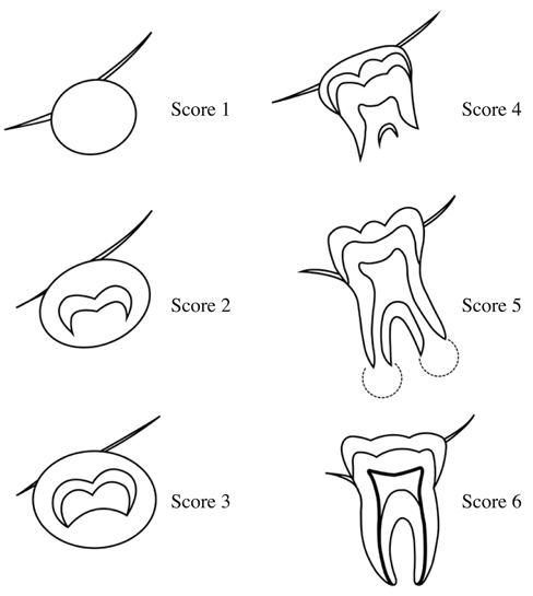
The first part of this study relied on measurements taken from panoramic radiographs of 614 Thai patients aged from 9 to 20. The stage of lower left and right third molar development was observed in each radiograph and a modified Gat score was assigned. Linear regression on this data produced the following equation: Y=9.309+1.673 mG+0.303S (Y=age; mG=modified Gat score; S=sex). In the second part of this study, the predictive accuracy of this equation was evaluated using data from a second set of panoramic radiographs (539 Thai subjects, 9 to 24 years old). Each subject's age was estimated using the above equation and compared against age calculated from a provided date of birth. Estimated and known age data were analyzed using the Pearson correlation coefficient and descriptive statistics.
RESULTS
Ages estimated from lower left and lower right third molar development stage were significantly correlated with the known ages (r=0.818, 0.808, respectively, P≤0.01). 50% of age estimates in the second part of the study fell within a range of error of ±1 year, while 75% fell within a range of error of ±2 years. The study found that the equation tends to estimate age accurately when individuals are 9 to 20 years of age.
CONCLUSION
The equation can be used for age estimation for Thai populations when the individuals are 9 to 20 years of age.
MeSH Terms
Figure
Reference
-
1. Crowder C, Austin D. Age ranges of epiphyseal fusion in the distal tibia and fibula of contemporary males and females. J Forensic Sci. 2005; 50:1001–1007.
Article2. Cameriere R, De Luca S, Biagi R, Cingolani M, Farronato G, Ferrante L. Accuracy of three age estimation methods in children by measurements of developing teeth and carpals and epiphyses of the ulna and radius. J Forensic Sci. 2012; 57:1263–1270.
Article3. Varshosaz M, Ehsani S, Nouri M, Tavakoli MA. Bone age estimation by cervical vertebral dimensions in lateral cephalometry. Prog Orthod. 2012; 13:126–131.
Article4. Lottering N, Macgregor DM, Meredith M, Alston CL, Gregory LS. Evaluation of the Suchey-Brooks method of age estimation in an Australian subpopulation using computed tomography of the pubic symphyseal surface. Am J Phys Anthropol. 2013; 150:386–399.
Article5. Wolff K, Vas Z, Sótonyi P, Magyar LG. Skeletal age estimation in Hungarian population of known age and sex. Forensic Sci Int. 2012; 223:374.e1–374.e8.
Article6. Halcrow SE, Tayles N, Buckley HR. Age estimation of children from prehistoric Southeast Asia: are the dental formation methods used appropriate? J Archaeol Sci. 2007; 34:1158–1168.
Article7. Kamalanathan GS, Hauck HM. Dental development of children in a Siamese village, Bang Chan, 1953. J Dent Res. 1960; 39:455–461.
Article8. Raungpaka S. The study of tooth-development age of Thai children in Bangkok. J Dent Assoc Thai. 1988; 38:72–81.9. Thevissen PW, Pittayapat P, Fieuws S, Willems G. Estimating age of majority on third molars developmental stages in young adults from Thailand using a modified scoring technique. J Forensic Sci. 2009; 54:428–432.
Article10. Krailassiri S, Anuwongnukroh N, Dechkunakorn S. Relationships between dental calcification stages and skeletal maturity indicators in Thai individuals. Angle Orthod. 2002; 72:155–166.11. Gat H, Sarnat H, Bjorvath K, Dayan D. Dental age evaluation. A new six-developmental-stage method. Clin Prev Dent. 1984; 6:18–22.12. Demirjian A, Goldstein H, Tanner JM. A new system of dental age assessment. Hum Biol. 1973; 45:211–227.13. Mincer HH, Harris EF, Berryman HE. The A.B.F.O. study of third molar development and its use as an estimator of chronological age. J Forensic Sci. 1993; 38:379–390.
Article14. Kullman L. Accuracy of two dental and one skeletal age estimation method in Swedish adolescents. Forensic Sci Int. 1995; 75:225–236.
Article15. Nykanen R, Espeland L, Kvaal SI, Krogstad O. Validity of the Demirjian method for dental age estimation when applied to Norwegian children. Acta Odontol Scand. 1998; 56:238–244.16. Uzamis M, Kansu O, Taner TU, Alpar R. Radiographic evaluation of third-molar development in a group of Turkish children. ASDC J Dent Child. 2000; 67:136–141.17. Bolanos MV, Moussa H, Manrique MC, Bolanos MJ. Radiographic evaluation of third molar development in Spanish children and young people. Forensic Sci Int. 2003; 133:212–219.18. Gunst K, Mesotten K, Carbonez A, Willems G. Third molar root development in relation to chronological age: a large sample sized retrospective study. Forensic Sci Int. 2003; 136:52–57.
Article19. Olze A, Taniguchi M, Schmeling A, Zhu BL, Yamada Y, Maeda H, et al. Comparative study on the chronology of third molar mineralization in a Japanese and a German population. Leg Med (Tokyo). 2003; 5:Suppl 1. S256–S260.
Article20. Mesotten K, Gunst K, Carbonez A, Willems G. Dental age estimation and third molars: a preliminary study. Forensic Sci Int. 2002; 129:110–115.
Article21. Willershausen B, Löffler N, Schulze R. Analysis of 1202 orthopantograms to evaluate the potential of forensic age determination based on third molar developmental stages. Eur J Med Res. 2001; 6:377–384.22. Darji JA, Govekar G, Kalele SD, Hariyani H. Age estimation from third molar development: A radiological study. J Indian Acad Forensic Med. 2011; 33:130–134.23. Bagherpour A, Anbiaee N, Partovi P, Golestani S, Afzalinasab S. Dental age assessment of young Iranian adults using third molars: a multivariate regression study. J Forensic Leg Med. 2012; 19:407–412.
Article24. Mahasantipiya PM, Pramojanee S, Thaiupathump T. Image analysis of the eruptive positions of third molars and adjacent second molars as indicators of age evaluation in Thai patients. Imaging Sci Dent. 2013; 43:289–293.
Article
- Full Text Links
- Actions
-
Cited
- CITED
-
- Close
- Share
- Similar articles
-
- Correlation of distal caries in the mandibular second molar and eruption state of the mandibular third molar
- A radiographic evaluation on the eruptional and positional changes of impacted mandibular third molars in young adults
- Correlation of pericoronitis and eruption state of the mandibular third molar
- Correlation of Left Mandibular Third Molar Development and Chronological Age
- The influence of mandibular third molar on mandibular angle fracture


