Comparison of transverse dental changes induced by the palatally applied Frog appliance and buccally applied Karad's integrated distalizing system
- Affiliations
-
- 1Department of Orthodontics, Faculty of Dentistry, Gazi University, Ankara, Turkey. fduzuner@yahoo.com.tr
- KMID: 2159357
- DOI: http://doi.org/10.4041/kjod.2016.46.2.96
Abstract
OBJECTIVE
To compare the transverse dental changes induced by the palatally applied Frog appliance and buccally applied Karad's integrated distalizing system (KIDS).
METHODS
We evaluated the pre- and post distalization orthodontic models of 39 patients, including 19 treated using the Frog appliance, which is palatally positioned (Frog group), and 20 treated using KIDS, which is buccally positioned (KIDS group). Changes in intermolar and interpremolar distances and the amount of maxillary premolar and molar rotation were evaluated on model photocopies. Wilcoxon and Mann-Whitney U tests were used for statistical evaluations. A p-value of < 0.05 was considered statistically significant.
RESULTS
Significant distopalatal rotation of premolars and distobuccal rotation of molars were observed in Frog group (p < 0.05), while significant distopalatal rotation of molars (p < 0.05), with no significant changes in premolars, was observed in KIDS group. The amount of second premolar and first molar rotation was significantly different between the two groups (p < 0.05 and p < 0.001, respectively). Furthermore, expansion in the region of the first molars and second premolars was significantly greater in KIDS group than in Frog group (p < 0.001 for both).
CONCLUSIONS
Our results suggest that the type and amount of first molar rotation and expansion vary with the design of the distalization appliance used.
Figure
-
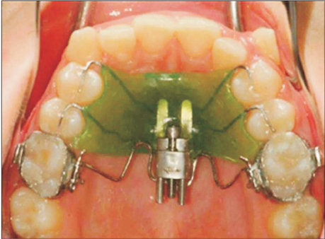
Figure 1 Frog appliance (Forestadent, Pfarzheim, Germany).
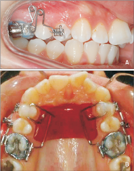
Figure 2 Karad's integrated distalizing system. A, Intraoral view from the buccal aspects. B, Intraoral view from the palatal aspects.
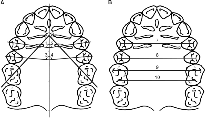
Figure 3 Measurements recorded on model photocopies. A, Angular measurements. 1, R4 angle: angle between the maxillary right first premolar axis and the midline reference plane (ML); 2, L4 angle: angle between the maxillary left first premolar axis and ML; 3, R5 angle: angle between the maxillary right second premolar axis and ML; 4, L5 angle: angle between the maxillary left second premolar axis and ML; 5, R6 angle: angle between the line passing through the distobuccal and mesiopalatal cusp tips of the maxillary right first molar and ML; 6, L6 angle: angle between the line passing through the distobuccal and mesiopalatal cusp tips of the maxillary left first molar and ML. B, Linear measurements. 7, U4 distance: distance between the buccal cusp tips of the maxillary right and left first premolars; 8, U5 distance: distance between the buccal cusp tips of the maxillary right and left second premolars; 9, U6M distance: distance between the mesiobuccal cusp tips of the maxillary right and left first molars; 10, U6D distance: distance between the distobuccal cusp tips of the maxillary right and left first molars.
Cited by 1 articles
-
Cone-beam computed tomography-guided three-dimensional evaluation of treatment effectiveness of the Frog appliance
Mujia Li, Xiaoxia Su, Yang Li, Xianglin Li, Xinqin Si
Korean J Orthod. 2019;49(3):161-169. doi: 10.4041/kjod.2019.49.3.161.
Reference
-
1. Papadopoulos MA. Orthodontic treatment of the Class II noncompliant patient. Current principles and techniques. Edinburgh: Mosby Elsevier;2006.2. Fontana M, Cozzani M, Caprioglio A. Noncompliance maxillary molar distalizing appliances: an overview of the last decade. Prog Orthod. 2012; 13:173–184.
Article3. Kinzinger GS, Eren M, Diedrich PR. Treatment effects of intraoral appliances with conventional anchorage designs for non-compliance maxillary molar distalization: a literature review. Eur J Orthod. 2008; 30:558–571.
Article4. Kinzinger GS, Fritz UB, Sander FG, Diedrich PR. Efficiency of a pendulum appliance for molar distalization related to second and third molar eruption stage. Am J Orthod Dentofacial Orthop. 2004; 125:8–23.
Article5. Mariani L, Maino G, Caprioglio A. Skeletal versus conventional intraoral anchorage for the treatment of class II malocclusion: dentoalveolar and skeletal effects. Prog Orthod. 2014; 15:43.
Article6. Grec RH, Janson G, Branco NC, Moura-Grec PG, Patel MP, Castanha Henriques JF. Intraoral distalizer effects with conventional and skeletal anchorage: a meta-analysis. Am J Orthod Dentofacial Orthop. 2013; 143:602–615.
Article7. Fontana M, Cozzani M, Mutinelli S, Spena R, Caprioglio A. Maxillary molar distalization therapy in adult patients: a multicentre study. Orthod Craniofac Res. 2015; 18:221–231.
Article8. Ludwig B, Glasl B, Kinzinger GS, Walde KC, Lisson JA. The skeletal frog appliance for maxillary molar distalization. J Clin Orthod. 2011; 45:77–84.9. Karad A. KIDS: a new approach to distalize maxillary molars. World J Orthod. 2008; 9:244–254.10. Braun S, Kusnoto B, Evans CA. The effect of maxillary first molar derotation on arch length. Am J Orthod Dentofacial Orthop. 1997; 112:538–544.
Article11. Korn M, Melsen B. Early treatment with a maxillary lip bumper-bite plateau combination. Angle Orthod. 2008; 78:838–846.
Article12. Hourfar J, Ludwig B, Kanavakis G. An active, skeletally anchored transpalatal appliance for derotation, distalization and vertical control of maxillary first molars. J Orthod. 2014; 41:Suppl 1. S24–S32.
Article13. Nalcaci R, Kocoglu-Altan AB, Bicakci AA, Ozturk F, Babacan H. A reliable method for evaluating upper molar distalization: Superimposition of three-dimensional digital models. Korean J Orthod. 2015; 45:82–88.
Article14. Maino G, Mariani L, Bozzo I, Maino G, Caprioglio A. Maxillary molar distalization with MGBM-system in class II malocclusion. J Orthod Sci. 2013; 2:101–108.
Article15. Kinzinger GS, Wehrbein H, Diedrich PR. Molar distalization with a modified pendulum appliance--in vitro analysis of the force systems and in vivo study in children and adolescents. Angle Orthod. 2005; 75:558–567.16. Kircelli BH, Pektaş ZO, Kircelli C. Maxillary molar distalization with a bone-anchored pendulum appliance. Angle Orthod. 2006; 76:650–659.17. Kinzinger GS, Gülden N, Yildizhan F, Diedrich PR. Efficiency of a skeletonized distal jet appliance supported by miniscrew anchorage for noncompliance maxillary molar distalization. Am J Orthod Dentofacial Orthop. 2009; 136:578–586.
Article18. Erverdi N, Koyutürk O, Küçükkeles N. Nickel-titanium coil springs and repelling magnets: a comparison of two different intra-oral molar distalization techniques. Br J Orthod. 1997; 24:47–53.
Article19. Bondemark L, Kurol J. Distalization of maxillary first and second molars simultaneously with repelling magnets. Eur J Orthod. 1992; 14:264–272.
Article20. Papadopoulos MA, Melkos AB, Athanasiou AE. Noncompliance maxillary molar distalization with the first class appliance: a randomized controlled trial. Am J Orthod Dentofacial Orthop. 2010; 137:586.e1–586.e13.
Article21. Acar AG, Gürsoy S, Dinçer M. Molar distalization with a pendulum appliance K-loop combination. Eur J Orthod. 2010; 32:459–465.
Article22. Bayram M, Nur M, Kilkis D. The frog appliance for upper molar distalization: a case report. Korean J Orthod. 2010; 40:50–60.
Article23. Champagne M. Reliability of measurements from photocopies of study models. J Clin Orthod. 1992; 26:648–650.24. Dahlberg G. Statistical methods for medical and biological students. New York: Interscience Publications;1940.25. Antonarakis GS, Kiliaridis S. Maxillary molar distalization with noncompliance intramaxillary appliances in Class II malocclusion. A systematic review. Angle Orthod. 2008; 78:1133–1140.
Article26. Burhan AS. Combined treatment with headgear and the Frog appliance for maxillary molar distalization: a randomized controlled trial. Korean J Orthod. 2013; 43:101–109.
Article27. Ghosh J, Nanda RS. Evaluation of an intraoral maxillary molar distalization technique. Am J Orthod Dentofacial Orthop. 1996; 110:639–646.
Article28. Cetlin NM, Ten Hoeve A. Nonextraction treatment. J Clin Orthod. 1983; 17:396–413.29. Fuziy A, Rodrigues de, Janson G, Angelieri F, Pinzan A. Sagittal, vertical, and transverse changes consequent to maxillary molar distalization with the pendulum appliance. Am J Orthod Dentofacial Orthop. 2006; 130:502–510.
Article30. Bolla E, Muratore F, Carano A, Bowman SJ. Evaluation of maxillary molar distalization with the distal jet: a comparison with other contemporary methods. Angle Orthod. 2002; 72:481–494.
- Full Text Links
-
- Actions
-
Cited
- CITED
-
- Close
- Share
-
- Similar articles
-
- The frog appliance for upper molar distalization: a case report
- Expansion of the mandibular arch using a trombone appliance
- Analysis of effects from usage of skeletal anchorage-assisted Pendulum appliance on vertical component of craniofacial structure
- Cone-beam computed tomography-guided three-dimensional evaluation of treatment effectiveness of the Frog appliance
- Surgery-first Approach for Facial Asymmetry with Transverse Discrepancy Using Hyrax-type Palatal Expansion Appliance
