An Intraosseous Epidermoid Cyst That Originated from the Nail Bed of Great Toe with Concurrent Joint Infection: A Case Report
- Affiliations
-
- 1Department of Orthopedic Surgery, Seoul National University Bundang Hospital, Seongnam, Korea. bc.jo81@gmail.com
- 2Department of Surgery, Seoul National University Bundang Hospital, Seongnam, Korea.
- KMID: 2158431
- DOI: http://doi.org/10.14193/jkfas.2016.20.1.50
Abstract
- We report on a rare case of an intraosseous epidermoid cyst in the distal phalanx of the great toe with concurrent infection in a 71-year-old woman with diabetes mellitus. The lesion was initially considered simple infectious arthritis and concomitant osteomyelitis in a patient with diabetes. However, after surgery, an intraosseous epidermoid cyst originating from the nail bed and involving the articular surface of the distal phalanx was detected. The epidermoid cyst may have contributed to the infectious arthritis in the interphalangeal joint. The lesion was treated via mass excision, arthrotomy, debridement, and intravenous antibiotics.
MeSH Terms
Figure
-
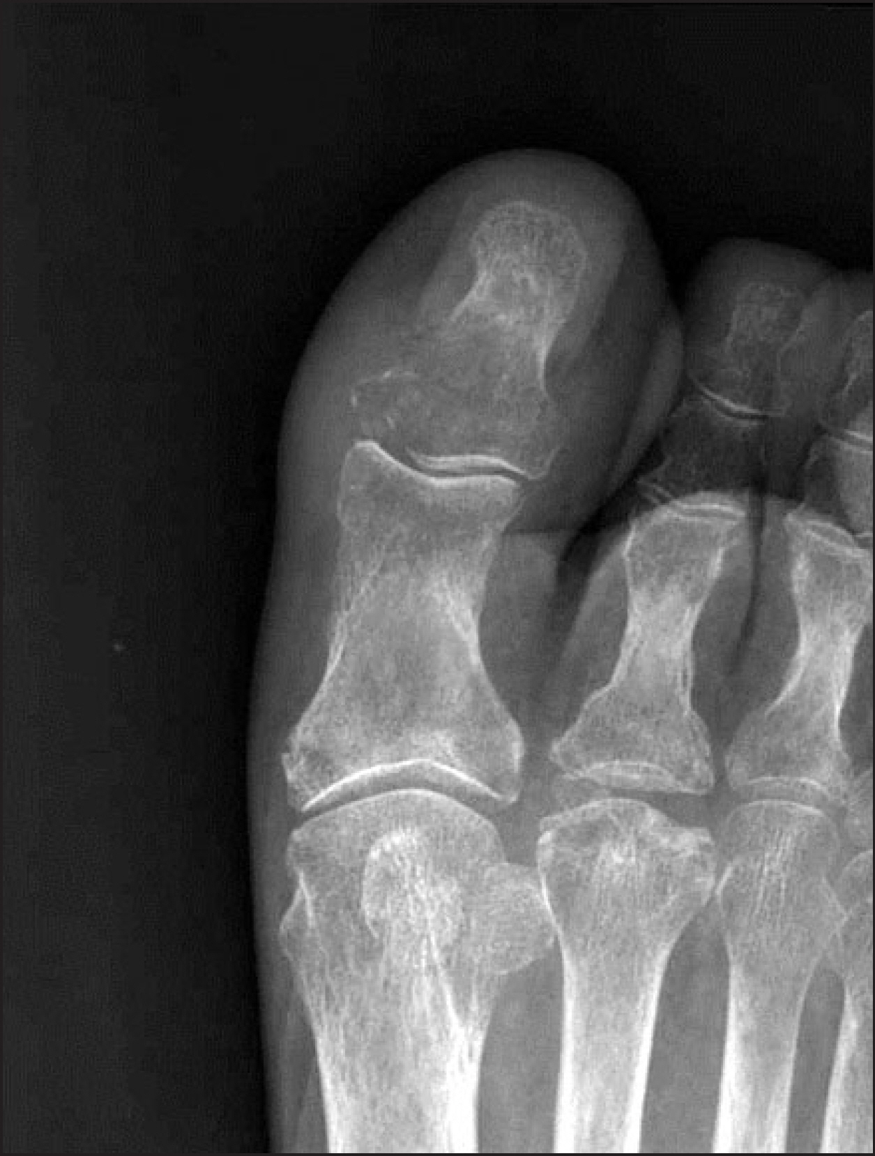
Figure 1. Preoperative anteroposterior radiograph reveals a radiolucent and osteolytic lesion at the base of the great toe's distal phalanx, which is more prominent in the medial aspect.
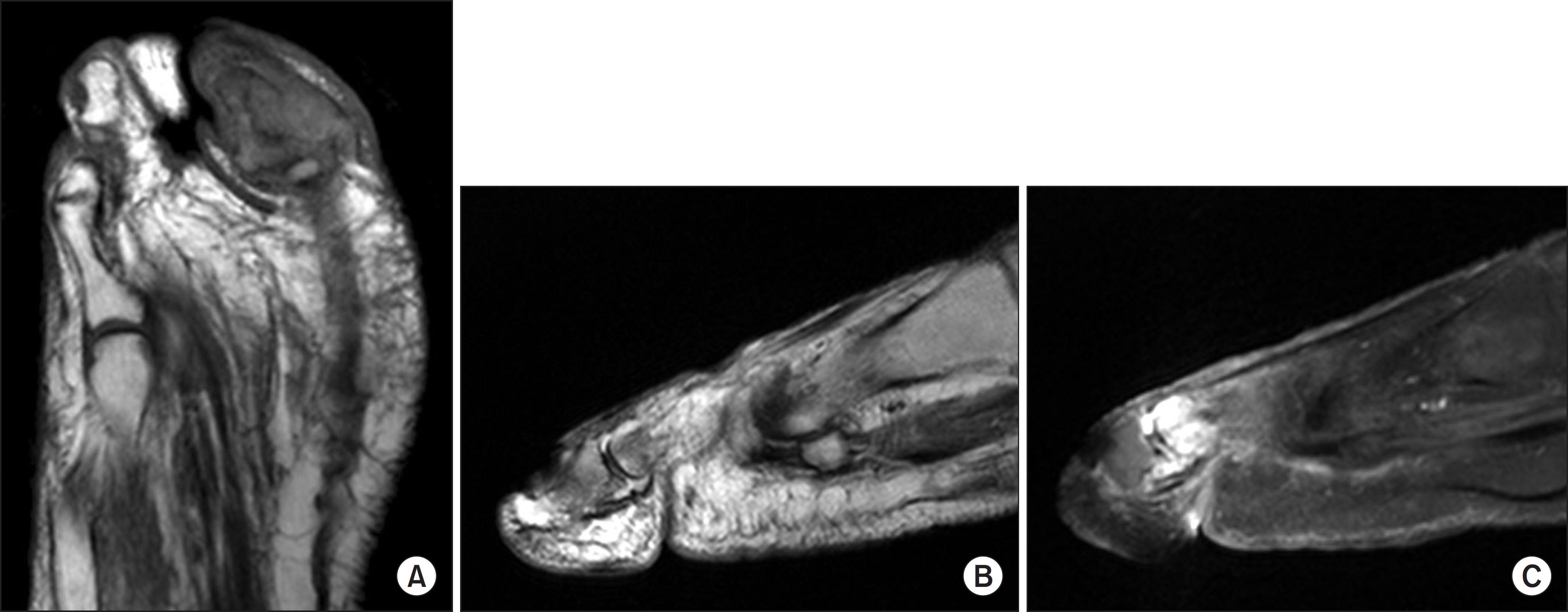
Figure 2. Preoperative magnetic resonance imaging findings. (A) A coronal T1-weighted image reveals an irregularly contoured nodular lesion at the distal phalanx. This lesion involves the great toe's distal phalanx with bony erosion and the adjacent flexor tendon. (B) A sagittal T2-weighted image reveals low signal intensity at the proximal and distal phalanx. (C) An enhanced T1-weighted image reveals low signal intensity at the great toe's proximal phalanx, synovial thickening with enhancement, and a small amount of joint effusion at the interphalangeal joint.

Figure 3. Intraoperative findings. (A) The extracted nail plate and attached intraosseous epidermoid cyst. A dirty creamy-brown material filled the cyst. (B) The defect in the medial side of the nail bed due to the intraosseous epidermoid cyst. Mass excision and curettage was performed throughout the defect, and a silastic drain was implanted.
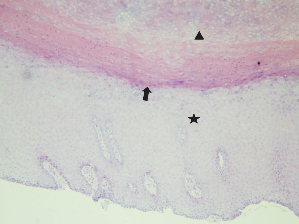
Figure 4. Histopathological findings (H&E stain, ×100). The intraosseous epidermoid cyst consists of the cyst wall (asterisk), granular layer (arrow), and copious keratin material (arrowhead). The cyst wall is lined with squamous cell epithelium and contains some skin appendages.
Reference
-
References
1. Patel K, Bhuiya T, Chen S, Kenan S, Kahn L. Epidermal inclusion cyst of phalanx: a case report and review of the literature. Skeletal Radiol. 2006; 35:861–3.
Article2. Schajowicz F, Aiello CL, Slullitel I. Cystic and pseudocystic lesions of the terminal phalanx with special reference to epidermoid cysts. Clin Orthop Relat Res. 1970; 68:84–92.3. Vion-Dury J, Vincentelli F, Jiddane M, Van Bunnen Y, Rumeau C, Grisoli F, et al. MR imaging of epidermoid cysts. Neuroradiology. 1987; 29:333–8.
Article4. Zadek I, Cohen HG. Epidermoid cyst of the terminal phalanx of a finger; with a review of the literature. Am J Surg. 1953; 85:771–4.5. Berlin SJ. A laboratory review of 67,000 foot tumors and lesions. J Am Podiatry Assoc. 1984; 74:341–7.
Article6. Mercuri M, Casadei R. Tumours in the foot. Foot Ankle Surg. 2002; 8:175–90.
Article7. Harris RI. Sebaceous cyst of terminal phalanx of thumb1 J Bone Joint Surg Am. 1930; 12:647–8.8. Connolly JE, Ratcliffe NR. Intraosseous epidermoid inclusion cyst presenting as a paronychia of the hallux. J Am Podiatr Med Assoc. 2010; 100:133–7.
Article
- Full Text Links
-
- Actions
-
Cited
- CITED
-
- Close
- Share
-
- Similar articles
-
- Intraosseous Epidermoid Cyst of Distal Phalanx: A case report
- A Case of Digital Intraosseous Epidermoid Inclusion Cyst of the Distal Phalanx
- A Case of Squamous Cell Carcinoma of the Nail Bed with Lymph Node Metastasis
- Thin Split-Thickness Toe Nail-Bed Grafts for Nail Bed Defects in Subungal Exostosis: Two Cases Report
- Tratment of the Nail Bed Avulsion Injury with Split-thickness Nail Bed Graft
