J Korean Foot Ankle Soc.
2016 Mar;20(1):27-31. 10.14193/jkfas.2016.20.1.27.
The Effect of Dwyer's Osteotomy and the 1st Metatarsal Osteotomy for Cavovarus Correction on Radiographic Parameters
- Affiliations
-
- 1W Institute for Foot and Ankle Disease and Trauma, W Hospital, Daegu, Korea.
- 2Department of Orthopedic Surgery, Ilsan Paik Hospital, Inje University College of Medicine, Goyang, Korea. sjs0506@paik.ac.kr
- KMID: 2158425
- DOI: http://doi.org/10.14193/jkfas.2016.20.1.27
Abstract
- PURPOSE
Several techniques have been introduced for correction of pes cavo-varus deformity. We retrospectively reviewed and compared the data of patients who underwent 1st metatarsal osteotomy alone, Dwyer's osteotomy alone, and 1st metatarsal osteotomy combined with Dwyer's osteotomy to determine the effect on radiographic parameters.
MATERIALS AND METHODS
Data on 28 cases in 27 consecutive patients recruited from 2006 to 2014 who underwent 1st metatarsal osteotomy alone (group F), Dwyer's osteotomy alone (group H), or 1st metatarsal osteotomy followed by Dwyer's osteotomy (group HF) with a minimum 1-year follow-up were reviewed retrospectively.
RESULTS
Calcaneal pitch angle on the standing foot lateral radiographs was significantly decreased after the operation in groups H and HF whereas Meary angle was decreased in groups F and HF. Hindfoot alignment angle and ratio on the hindfoot alignment view were improved in groups H and HF. Maximal medial cuneiform height reduction was observed in group HF. 1st ray was significantly shortened in groups F and HF.
CONCLUSION
Combined forefoot and hindfoot operation took the largest correction power of all radiologic parameters.
Keyword
MeSH Terms
Figure
-
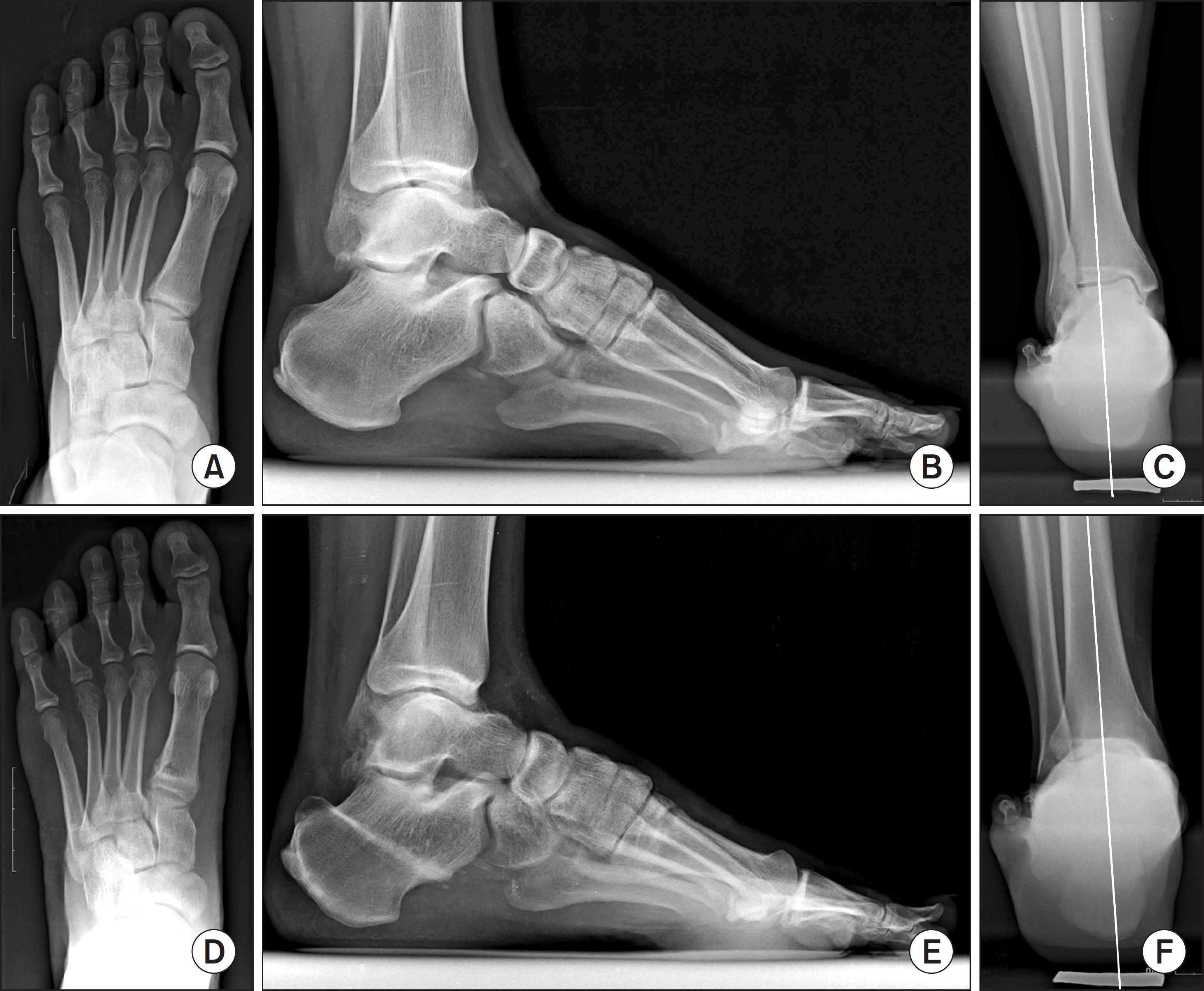
Figure 1. (A∼C) Preoperative radiographs show increased Meary angle (16o) calcaneal pitch angle (33o) and hindfoot varus. (D∼F) Postoperative radiographs show improvement of Meary angle (8o) and calcaneal pitch angle (24o) and hindfoot varus with simultaneous forefoot and hindfoot correction.
Reference
-
References
1. Coughlin MJ, Mann RA. Pes cavus1 In: Coughlin MJ, Mann RA, Saltzmann CL, editors. Mann's surgery of the foot and ankle. 9th ed.Philadelphia: Mosby;2014. p. 11361–82.2. Steindler A. Operative treatment of pes cavus: stripping of the os calcis. Surg Gynecol Obstet. 1917; 24:612–5.3. Coughlin MJ. Lesser toe abnormalities. J Bone Joint Surg Am. 2002; 84:1446–69.4. Coughlin MJ. Subluxation and dislocation of the second metatarsophalangeal joint. Orthop Clin North Am. 1989; 20:535–51.5. Knupp M, Horisberger M, Hintermann B. A new z-shaped calcaneal osteotomy for 3-plane correction of severe varus deformity of the hindfoot. Tech Foot Ankle Surg. 2008; 7:90–5.
Article6. Coleman SS, Chesnut WJ. A simple test for hindfoot flexibility in the cavovarus foot. Clin Orthop Relat Res. 1977; 123:60–2.
Article7. Saltzman CL, el-Khoury GY. The hindfoot alignment view. Foot Ankle Int. 1995; 16:572–6.
Article8. Lee HS, Wapner KL, Park SS, Kim JS, Lee DH, Sohn DW. Ligament reconstruction and calcaneal osteotomy for osteoarthritis of the ankle. Foot Ankle Int. 2009; 30:475–80.
Article9. Kitaoka HB, Alexander IJ, Adelaar RS, Nunley JA, Myerson MS, Sanders M. Clinical rating systems for the ankle-hindfoot, midfoot, hallux, and lesser toes. Foot Ankle Int. 1994; 15:349–53.
Article10. Benzton PGK. Pes cavus and the M. peroneus longus. Acta Orthop Scand. 1933; 4:50–2.11. Halgrimsson S. Pes cavus, seine Behandlung und Einige Be-merkungen uber seing Aetiologie [Pes cavus, its treatment and some remarks about its etiology]. Acta Orthop Scand. 1939; 10:73–1181. German.12. Duchenne GB. The physiology of motion (trans EB Kaplan). Philadelphia: WB Saunders;1959.13. Nogueira MP, Farcetta F, Zuccon A. Cavus foot. Foot Ankle Clin1. 2015; 20:645–56.
Article14. Myerson MS, Shereff MJ. The pathological anatomy of claw and hammer toes. J Bone Joint Surg Am. 1989; 71:45–9.
Article15. Dwyer FC. Osteotomy of the calcaneum for pes cavus. J Bone Joint Surg Br. 1959; 41:80–6.
Article16. Tennant JN, Carmont M, Phisitkul P. Calcaneus osteotomy. Curr Rev Musculoskelet Med. 2014; 7:271–6.
Article17. Lamm BM, Gesheff MG, Salton HL, Dupuis TW, Zeni F. Preoperative planning and intraoperative technique for accurate realignment of the Dwyer calcaneal osteotomy. J Foot Ankle Surg. 2012; 51:743–8.
Article18. Kraus JC, Fischer MT, McCormick JJ, Klein SE, Johnson JE. Geometry of the lateral sliding, closing wedge calcaneal osteotomy: review of the two methods and technical tip to minimize shortening. Foot Ankle Int. 2014; 35:238–42.19. Zhou Y, Zhou B, Liu J, Tan X, Tao X, Chen W, et al. A prospective study of midfoot osteotomy combined with adjacent joint sparing internal fixation in treatment of rigid pes cavus deformity. J Orthop Surg Res. 2014; 9:44.
Article20. Bariteau JT, Blankenhorn BD, Tofte JN, DiGiovanni CW. What is the role and limit of calcaneal osteotomy in the cavovarus foot? Foot Ankle Clin. 2013; 18:697–714.
Article21. Faldini C, Traina F, Nanni M, Mazzotti A, Calamelli C, Fabbri D, et al. Surgical treatment of cavus foot in Charcot-Marie-tooth disease: a review of twenty-four cases: AAOS exhibit selection. J Bone Joint Surg Am. 2015; 97:e30.
- Full Text Links
-
- Actions
-
Cited
- CITED
-
- Close
- Share
-
- Similar articles
-
- Combined First Metatarsal and Calcaneal Osteotomy for Fixed Cavovarus Deformity of The Foot
- Comparison of Proximal Metatarsal Osteotomy andDistal Chevron Osteotomy for Correction of Hallux Valgus
- Treatment Results of Hallux Valgus Deformity by Parallel-Shaped Modified Scarf Osteotomy
- A Comparison of Operative Treatment of Hallux Valgus with a Proximal Metatarsal Osteotomy and with a Modified Chevron Osteotomy
- Comparison of the Results between Distal Chevron Osteotomy and Proximal Metatarsal Osteotomy for the Treatment of Moderate Hallux Valgus
