Paradoxical Heart Failure Precipitated by Profound Dehydration: Intraventricular Dynamic Obstruction and Significant Mitral Regurgitation in a Volume-Depleted Heart
- Affiliations
-
- 1Cardiology Division, Department of Internal Medicine, Dankook University College of Medicine, Cheonan, Korea.
- 2Presbyterian Medical Center, Jeonju, Korea.
- 3Department of Radiology, Gachon University of Medicine and Science, Incheon, Korea.
- 4Division of Cardiology, Department of Internal Medicine, Gachon University of Medicine and Science, Incheon, Korea. jeff76@gilhospital.com
- KMID: 2158246
- DOI: http://doi.org/10.3349/ymj.2013.54.4.1058
Abstract
- Occurrence of dynamic left ventricular outflow tract (LVOT) obstruction is not infrequent in critically ill patients, and it is associated with potential danger. Here, we report a case of transient heart failure with hemodynamic deterioration paradoxically induced by extreme dehydration. This article describes clinical features of the patient and echocardiographic findings of dynamic LVOT obstruction and significant mitral regurgitation caused by systolic anterior motion of the mitral valve in a volume-depleted heart.
MeSH Terms
Figure
-
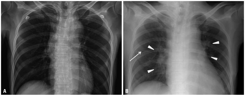
Fig. 1 (A) Chest radiograph taken previously. (B) Chest radiograph showing newly developed pulmonary congestion (arrow heads) and fluid collection in the fissure (arrow) at the time of presentation.
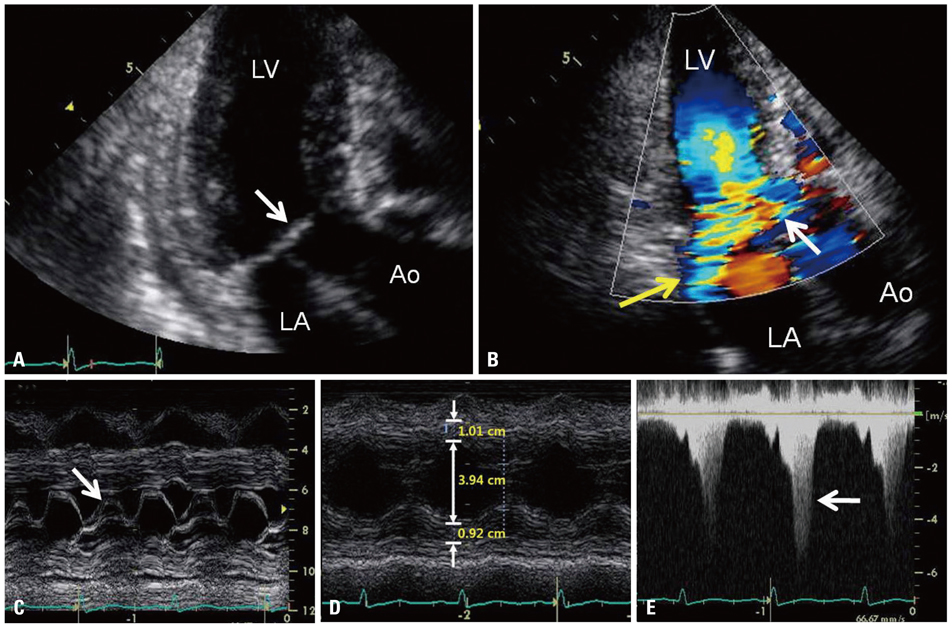
Fig. 2 (A) Two-dimensional transthoracic echocardiography shows systolic anterior motion of the mitral leaflets (arrow) with left ventricular outflow tract obstruction. (B) Color Doppler image reveals flow acceleration at the left ventricular outflow tract (white arrow) and significant mitral regurgitation (yellow arrow). (C) M-mode image shows systolic anterior motion of the mitral valve leaflets (arrow). (D) M-mode image at the midventricular cavity shows small left ventricular cavity and normal wall thickness.
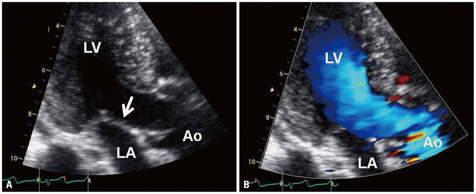
Fig. 3 (A) Two-dimensional transthoracic echocardiography reveals disappearance of systolic anterior motion of mitral leaflets (arrow) after the patient's condition had been stabilized. (B) Color flow image shows resolution of flow acceleration and normal lamina flow at the left ventricular outflow tract. Concurrently, this color flow image after volume restoration therapy shows that previously observed mitral regurgitation has been completely abolished. LA, left atrium; LV, left ventricle; Ao, ascending aorta.
Reference
-
1. Haley JH, Sinak LJ, Tajik AJ, Ommen SR, Oh JK. Dynamic left ventricular outflow tract obstruction in acute coronary syndromes: an important cause of new systolic murmur and cardiogenic shock. Mayo Clin Proc. 1999; 74:901–906.
Article2. Aurigemma G, Battista S, Orsinelli D, Sweeney A, Pape L, Cuénoud H. Abnormal left ventricular intracavitary flow acceleration in patients undergoing aortic valve replacement for aortic stenosis. A marker for high postoperative morbidity and mortality. Circulation. 1992; 86:926–936.
Article3. Villareal RP, Achari A, Wilansky S, Wilson JM. Anteroapical stunning and left ventricular outflow tract obstruction. Mayo Clin Proc. 2001; 76:79–83.
Article4. Mingo S, Benedicto A, Jimenez MC, Pérez MA, Montero M. Dynamic left ventricular outflow tract obstruction secondary to catecholamine excess in a normal ventricle. Int J Cardiol. 2006; 112:393–396.
Article5. Yang JH, Park SW, Yang JH, Cho SW, Kim HS, Choi KA, et al. Dynamic left ventricular outflow tract obstruction without basal septal hypertrophy, caused by catecholamine therapy and volume depletion. Korean J Intern Med. 2008; 23:106–109.
Article6. Chockalingam A, Dorairajan S, Bhalla M, Dellsperger KC. Unexplained hypotension: the spectrum of dynamic left ventricular outflow tract obstruction in critical care settings. Crit Care Med. 2009; 37:729–734.
Article7. Verbalis JG. Disorders of body water homeostasis. Best Pract Res Clin Endocrinol Metab. 2003; 17:471–503.
Article8. Topalian S, Ginsberg F, Parrillo JE. Cardiogenic shock. Crit Care Med. 2008; 36:1 Suppl. S66–S74.
Article
- Full Text Links
-
- Actions
-
Cited
- CITED
-
- Close
- Share
-
- Similar articles
-
- Percutaneous Edge-to-Edge Mitral Valve Repair for Functional Mitral Regurgitation
- Common Co-Morbidities in Heart Failure – Diabetes, Functional Mitral Regurgitation and Sleep Apnoea
- Echocardiographic Assessment of Mitral Valve Regurgitation
- The Unusual Suspect: Anemia-induced Systolic Anterior Motion of the Mitral Valve and Intraventricular Dynamic Obstruction in a Hyperdynamic Heart as Unexpected Causes of Exertional Dyspnea after Cardiac Surgery
- Acute Mitral Regurgitation due to Spontaneous Chordal Rupture in a Patient With Obstructive Hypertrophic Cardiomyopathy
