Yonsei Med J.
2007 Aug;48(4):715-718. 10.3349/ymj.2007.48.4.715.
Spontaneous Regression of a Cystic Tumor in a Postpartum Woman; Is It A Cystic Lymphangioma?
- Affiliations
-
- 1Department of Diagnostic Radiology, Yonsei University College of Medicine, Seoul, Korea. kimnex@yuhs.ac
- 2Department of Surgery, Yonsei University College of Medicine, Seoul, Korea.
- 3Brain Korea 21 Project for Medical Science, Institute of Gastroenterology, Yonsei University College of Medicine, Seoul, Korea.
- KMID: 2158181
- DOI: http://doi.org/10.3349/ymj.2007.48.4.715
Abstract
- Spontaneous regression of intra-abdominal cystic tumors in adults is unusual. Here, we present the case of an apparently spontaneous regression of a large intra-abdominal cystic mass found in the postpartum period of an 18-year-old woman. The regression was demonstrated using serial computed tomography (CT) examinations over a two-year period.
Keyword
MeSH Terms
Figure
-
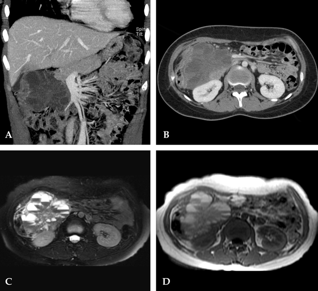
Fig. 1 Coronal (A) and axial (B) view of contrast enhanced CT scan demonstrating a large, multilobulated, multilocular, heterogeneous cystic mass with internal septa in the right upper quadrant abdomen. On axial T2-weighted spin echo image (8000/105) with fat suppression (C) and axial T1-weighted incoherent gradient-echo (SPGR) images (230/4.2) (D), the signal intensity of the upper portions of the fluid within the cysts showed high signal intensity, and the area of the lower portions of fluid showed low signal intensity.
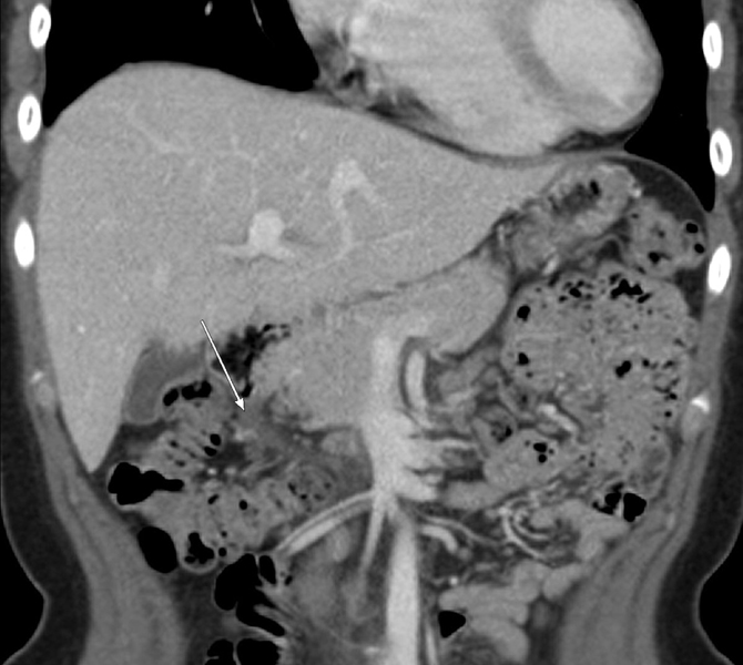
Fig. 2 Follow-up CT scan two years after initial examination shows marked regression of the mass with a tiny remaining cystic lesion in the right upper quadrant abdomen.
Reference
-
1. Chen JS, Lee WJ, Chang YJ, Wu MZ, Chiu KM. Laparoscopic resection of a primary retroperitoneal mucinous cystadenoma: report of a case. Surg Today. 1998. 28:343–345.
Article2. Ros PR, Olmsted WW, Moser RP, Dachman AH, Hjermstad BH, Sobin LH. Mesenteric and omental cysts: histologic classification with imaging correlation. Radiology. 1987. 164:327–332.
Article3. Stoupis C, Ros PR, Abbitt PL, Burton SS, Gauger J. Bubbles in the belly: imaging of cystic mesenteric or omental masses. Radiographics. 1994. 14:729–737.
Article4. Yang DM, Jung DH, Kim H, Kang JH, Kim SH, Kim JH, et al. Retroperitoneal cystic masses: CT, clinical, and pathologic findings and literature review. Radiographics. 2004. 24:1353–1365.
Article5. Davidson AJ, Hartman DS. Lymphangioma of the retroperitoneum: CT and sonographic characteristic. Radiology. 1990. 175:507–510.
Article6. Choi JY, Kim MJ, Chung JJ, Park SI, Lee JT, Yoo HS, et al. Gallbladder lymphangioma: MR findings. Abdom Imaging. 2002. 27:54–57.
Article7. Liew SC, Glenn DC, Storey DW. Mesenteric cyst. Aust N Z J Surg. 1994. 64:741–744.
Article8. Torashima Y, Yamaguchi J, Taniguchi K, Fujioka H, Shimokawa I, Izawa K, et al. Surgery for ileal mesenteric lymphangioma during pregnancy: case report and review of the literature. J Gastrointest Surg. 2004. 8:616–620.
Article9. Quack Loetscher KC, Jandali AR, Garzoli E, Pok J, Beinder E. Axillary cavernous lymphangioma in pregnancy and puerperium. Gynecol Obstet Invest. 2005. 60:108–111.
Article10. Warner AA, Kofinas AD, Melone PJ. Spontaneous resolution of cystic hygroma. Prenat Diagn. 1990. 10:758.
Article11. Distell BM, Hertzberg BS, Bowie JD. Spontaneous resolution of a cystic neck mass in a fetus with normal karyotype. AJR Am J Roentgenol. 1989. 153:380–382.
Article12. Watson WJ, Katz VL, Thorp JM. Spontaneous resolution of fetal nuchal cystic hygroma. J Perinatol. 1991. 11:213–215.13. Wu MP, Wu RC, Lee JS, Yao WJ, Kuo PL. Spontaneous resolution of fetal mediastinal cystic hygroma. Int J Gynaecol Obstet. 1995. 48:295–298.
Article14. de Perrot M, Bründler M, Tötsch M, Mentha G, Morel P. Mesenteric cysts. Toward less confusion? Dig Surg. 2000. 17:323–328.15. Takiff H, Calabria R, Yin L, Stabile BE. Mesenteric cysts and intra-abdominal cystic lymphangiomas. Arch Surg. 1985. 120:1266–1269.
Article16. Siegel MJ, Glazer HS, St Amour TE, Rosenthal DD. Lymphangiomas in children: MR imaging. Radiology. 1989. 170:467–470.
Article17. Ozgen A, Akata D, Akhan O, Tez M, Gedikoglu G, Ozmen MN. Giant benign cystic peritoneal mesothelioma: US, CT, and MRI findings. Abdom Imaging. 1998. 23:502–504.18. Bos SD, Jansen W, Ypma AF. Multicystic mesothelioma presenting as a pelvic tumour: case report and literature review. Scand J Urol Nephrol. 1995. 29:225–228.
Article19. Hornick JL, Fletcher CD. Intraabdominal cystic lymphangiomas obscured by marked superimposed reactive changes: clinicopathological analysis of a series. Hum Pathol. 2005. 36:426–432.
Article20. Daniel S, Lazarevic B, Attia A. Lymphangioma of the mesentery of the jejunum: report of a case and a brief review of the literature. Am J Gastroenterol. 1983. 78:726–729.21. Maniwa Y, Tsubota N, Yoshimura M, Murotani A, Miyamoto Y, Takagi Y, et al. A case of the localized fibrous mesothelioma which size decreased temporarily. Kyobu Geka. 1994. 47:948–951.22. Robinson BW, Robinson C, Lake RA. Localised spontaneous regression in mesothelioma-possible immunological mechanism. Lung Cancer. 2001. 32:197–201.
Article23. Iyer R, Eftekhari F, Varma D, Jaffe N. Cystic retroperitoneal lymphangioma: CT, ultrasound and MR findings. Pediatr Radiol. 1993. 23:305–306.
Article
- Full Text Links
-
- Actions
-
Cited
- CITED
-
- Close
- Share
-
- Similar articles
-
- Cystic lymphangioma of the pancreas: a case report
- Retroperitoneal cystic lymphangioma in an aged man: report of a case and review of the literature
- Cystic Lymphangioma of the Breast in an Adult Woman
- Cystic Lymphangioma Involving the Mesentery and the Retroperitoneum: A Case Report
- Laparoscopic Excision of a Retroperitoneal Cystic Lymphangioma Encasing the Renal Vessels
