Littoral Cell Angioma (LCA) Associated with Liver Cirrhosis
- Affiliations
-
- 1Department of Internal Medicine, Inha University, Incheon, Korea. ywshin@inha.ac.kr
- 2Department of Pathology, Inha University, Incheon, Korea.
- 3Department of Surgery, Inha University, Incheon, Korea.
- KMID: 2158134
- DOI: http://doi.org/10.3349/ymj.2005.46.1.184
Abstract
- A littoral cell angioma (LCA) is a rare benign vascular tumor of the spleen. A 60-year-old man, with multiple nodules in imaging study and liver cirrhosis graded as Child-Pugh classification class A, was transferred for splenomegaly. A thrombocytopenia was found on hematological evaluation. Because there was no evidence of hematological and visceral malignancy, a splenectomy was performed for a definitive diagnosis. The histological and immunohistochemical features of the splenic specimens were consistent with a LCA. After the splenectomy, the thrombocytopenia recovered to the normal platelet count. There has been no previous report of a LCA combined with liver cirrhosis. Herein, the first case of a LCA in Korea, diagnosed and treated by a splenectomy, is reported.
Keyword
MeSH Terms
Figure
-
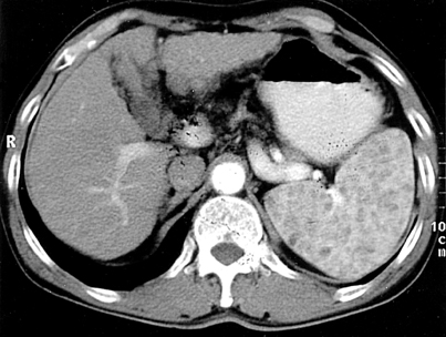
Fig. 1 Enhanced abdominal CT scan shows an enlarged spleen with multiple low attenuated nodular lesions and liver cirrhosis featuring a nodular surface, heterogeneous density and mild atrophy of the left lobe.
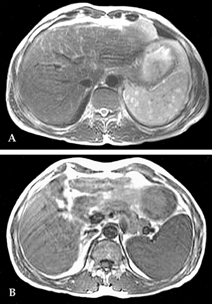
Fig. 2 A. T2-weighted abdominal magnetic resonance image shows multiple splenic nodules with high signal intensity. B. T1-weighted abdominal magnetic resonance image shows multiple splenic nodules with iso to low-signal intensity.
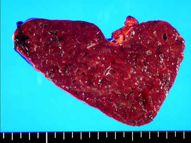
Fig. 3 The multiple circumscribed blood-filled spongy nodules of 0.2 to 0.6 cm in size located within the spleen that weighed 370 g. The capsule was intact and tensile, with a vaguely micronodular surface. The cut surface of the spleen was a dark purple color and beef-like soft.
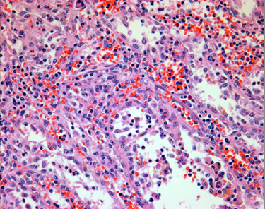
Fig. 4 Anastomosing vascular channels with a pseudopapillary pattern and exfoliated cells present in the luminal spaces of the vascular channels (H & E stain, × 400).

Fig. 5 Immunohistochemical staining of the splenic nodules. A. Positive staining for the Factor VIII. B. Positive staining for CD31. C. Positive staining for CD68. D. Negative staining for CD34.
Reference
-
1. Falk S, Stutte HJ, Frizzera G. Littoral cell angioma: A novel splenic vascular lesion demonstrating histologic differentiation. Am J Surg Pathol. 1991. 15:1023–1033.2. Rosso R, Paulli M, Gianelli U, Boveri E, Stella G, Magrini U. Littoral cell angiosarcoma of the spleen: Case report with immunohistochemical and ultrastructural analysis. Am J Surg Pathol. 1995. 19:1203–1208.3. Sauer J, Treichel U, Kohler HH, Schunk K, Junginger T. Littoral cell angioma: A rare differential diagnosis on splenic tumor. Dtsch Med Wochenschr. 1999. 24:624–628.4. Goldfeld M, Cohen I, Loberant N, Magrabi A, Katz I, Papura S, et al. Littoral cell angioma of the spleen: Appearance on sonography and CT. J Clin Ultrasound. 2002. 30:510–513.5. Ben-Izhak O, Bejar J, Ben-Elieezer S, Vlodavsky E. Splenic Littoral cell hemangioendothelioma: A new low-grade variant of malignant Littoral cell tumor. Histopathology. 2001. 369:469–475.6. Ziske C, Meybehm M, Sauerbruch T. Littoral cell angioma as a rare cause of splenomegaly. Ann Hematol. 2001. 80:45–48.7. Arber DA, Strickler JG, Chen YY, Weiss LM. Splenic vascular tumors: A histologic immunophenotypic and virologic study. Am J Surg Pathol. 1997. 21:827–835.8. Oliver-Goldaracena JM, Blanco A, Miralles M, Martin-Gonzales M. Littoral cell angioma of the spleen. Abdom Imaging. 1998. 23:636–639.9. Kinoshita LL, Yee J, Nash SR. Littoral cell angioma of the spleen: Imaging feature. Am J Roentgenol. 2000. 174:467.10. Levy AD, Abbott RM, Abbondanzo SL. Littoral cell angioma of the spleen: CT features with clinicopathologic comparision. Radiology. 2004. 230:485–490.11. Schneider G, Uder M, Altmeyer K, Bonkhoff H, Grube M, Kramann B. Littoral cell angioma of the spleen: CT and MR imaging appearance. Eur Radiol. 2000. 10:1395–1400.12. Heese J, Bocklage T. Specimen fine-needle aspiration cytology of littoral cell angioma with histologic and immunohistochemical confirmation. Diagn Cytopathol. 2000. 22:39–44.13. Lucey BC, Boland GW, Maher MM, Hahn PF, Gervais DA, Mueller PR. Percutaneous nonvascular splenic intervention : A 10-year review. Am J Roentgenol. 2002. 179:1591–1596.14. Keogan MT, Freed KS, Paulson EK, Nelson RC, Dodd LG. Imaging-guided percutaneous biopsy of focal splenic lesions: Update on safety and effectiveness. Am J Roentgenol. 1999. 172:933–937.15. Fishman EK, Magid D, Kuhlman JE. Pneumocystis carinii involvement of the liver and spleen: CT demonstration. J Comput Assist Tomogr. 1990. 14:146–148.16. Radin DR. Intraabdominal Mycobacterium tuberculosis vs Mycobacterium avium-intracellulare infections in patients with AIDS: Distinction based on CT findings. Am J Roentgenol. 1991. 156:487–491.17. Collins GL, Morgan MB, Tylor FM 3rd. Littoral cell angiomatosis with poorly differentiated adenocarcinoma of the lung. Ann Diagn Pathol. 2003. 7:54–59.18. Bisceglia M, Sickel J, Giangaspero F, et al. Littoral cell angioma of the spleen: An additional report of four cases with emphasis on the association with visceral organ cancer. Tumori. 1998. 84:595–599.
- Full Text Links
-
- Actions
-
Cited
- CITED
-
- Close
- Share
-
- Similar articles
-
- Splenic Littoral Cell Angioma with Hepatitis C Associated Liver Cirrhosis
- A Case of Bicytopenia Combined with Littoral Cell Angioma
- Littoral cell angiomas: Benign lesion with a penchant for visceral malignancies
- Hypothalamic-Pituitary-Gonadal Function in Hepatic Failure, Clinical Study
- Multiple Spider Angiomas as a Predictive Diagnostic Marker of Alcoholic Liver Cirrhosis
