Giant Fibrovascular Polyp of the Hypopharynx: Surgical Treatment with the Biappoach
- Affiliations
-
- 1Department of Thoracic and Cardiovascular Surgery, Sungkyunkwan University School of Medicine, Samsung Medical Center, Seoul, Korea. ymshim@smc.samsung.co.kr
- KMID: 2157833
- DOI: http://doi.org/10.3346/jkms.2006.21.4.749
Abstract
- Fibrovascular polyps of the esophagus and hypopharynx are benign tumors of the upper digestive tract. The majority of these polyps are located in the upper part of the esophagus but the hypopharyngeal fibrovascular polyps are only rarely seen. Most of them are surgically treated and this is usually done through a cervical incision, although some of them have been removed endoscopically. The authors report here on a case of a 63-yr-old-man with a giant fibrovascular polyp of the hypopharynx that extended into the stomach; this polyp was removed through simultaneous transcervical and transabdominal approaches because of the huge size of the polyp. The man presented with progressive dysphagia of 1 yr duration. The preoperative assessment revealed a giant polyp arising from the left arytenoid and extending into the stomach. The dimension of the polyp was about 26 x10 x4 cm. The complete resection of the polyp with the simultaneous transcervical and transabdominal approaches was successful, and it was diagnosed as a fibrovascular polyp. The patient has been followed up without any recurrence for 6 month postoperatively.
Keyword
MeSH Terms
Figure
-
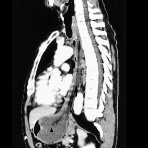
Fig. 1 Sagittal view of a chest computed tomography scan of a 63-yr-old-man with a giant fibrovascular polyp. A soft tissue lesion (arrow) in the esophagus from the level of the cervical esophagus extends to the level of the upper body of the stomach. The exact location of the polyp's origin is obscure. The dimension of the head (arrowhead) is about 8×5 cm in size. The central portion of the lesion shows attenuation identical to that of subcutaneous fat.

Fig. 2 Esophagoscopic photograph at the level of the hypopharynx. The base of the stalk (arrowhead) originates from the left arytenoid (arrow). PPW, posterior pharyngeal wall.

Fig. 3 The gross appearance of the giant fibrovascular polyp. The dimension of the polyp is about 26×10×4 cm. The polyp is covered with mucous membrane similar to that of the esophagus.
Cited by 1 articles
-
A Case of Large Fibrovascular Polyp of the Stomach
Eun Ji Lee, Seung Goun Hong, Hae Ri Baek, Chan Bok Lee, Sang Myung Choi, Sung Jin Kim, Byoung Gy Chae, Cheul Young Choi
Clin Endosc. 2013;46(2):186-188. doi: 10.5946/ce.2013.46.2.186.
Reference
-
1. Rice TW, Murthy SC. Sellke FW, del Nido PJ, Swanson SJ. Surgical treatment of benign esophageal diseases. Sabiston & Spencer Surgery of the Chest. 2005. Volume I:7th ed. Philadelphia: Elsevier Saunders;583–609.
Article2. Shamji F, Todd TR. Pearson FG, Cooper JD, Deslauriers J, Ginsberg RJ, Hiebert CA, Patterson GA, Urschel HC, editors. Benign tumors. Esophageal Surgery. 2002. 2nd ed. Philadelphia: Churchill Livingstone;637–654.3. Seshul MJ, Wiatrak BJ, Galliani CA, Odrezin GT. Pharyngeal fibrovascular polyp in a child. Ann Otol Rhinol Laryngol. 1998. 107:797–800.
Article4. Paik HC, Han JW, Jung EK, Bae KM, Lee YH. Fibrovascular polyp of the esophagus in infant. Yonsei Med J. 2001. 42:264–266.
Article5. Jang GC, Clouse ME, Fleischner FG. Fibrovascular polyp: a benign intraluminal tumor of the esophagus. Radiology. 1969. 92:1196–1200.6. Nuyens MR, Bhatti NI, Flint P. Multiple synchronous fibrovascular polyps of the hypopharynx. J Otorhinolaryngol Relat Spec. 2004. 66:341–344.
Article7. Avezzano EA, Fleischer DE, Merida MA, Anderson DL. Giant fibrovascular polyps of the esophagus. Am J Gastroenterol. 1990. 85:299–302.8. Devereaux BM, LeBlanc JK, Kesler K, Brooks J, Lehman GA, Sherman S, Ciaccia D. Giant fibrovascular polyp of the esophagus. Endoscopy. 2003. 35:970–972.
Article9. Schuhmacher C, Becker K, Dittler HJ, Hofler H, Siewert JR, Stein HJ. Fibrovascular esophageal polyp as a diagnostic challenge. Dis Esophagus. 2000. 13:324–327.
Article10. Marcial-Rojas RA, Suan P. Epidermoid carcinoma in mucosa overlying a pedunculated lipoma of the esophagus. J Thorac Surg. 1959. 37:427–434.
Article11. Tasaka Y, Makimoto K, Yamauchi M, Haebara H. Benign pedunculated intraluminal tumor of the esophagus. J Otolaryngol. 1982. 11:111–115.12. van Lanschot JJ, Poublon RM, Zonderland HM, van Houten H. Benign pedunculated tumor of the esophagus. Neth J Surg. 1987. 39:83–85.13. Oh SS, Shim YM. Giant fibrovascular polyp of the esophagus: a case report. Korean J Thorac Cardiovasc Surg. 1996. 29:675–680.14. Drenth J, Wobbes T, Bonenkamp JJ, Nagengast FM. Recurrent esophageal fibrovascular polyps: case history and review of the literature. Dig Dis Sci. 2002. 47:2598–2604.
- Full Text Links
-
- Actions
-
Cited
- CITED
-
- Close
- Share
-
- Similar articles
-
- A Case of Giant Fibrovascular Polyp of the Hypopharynx Removed by Transoral Approach
- Giant Fibrovascular Polyp of the Esophagus: A Case Report
- A Case of Fibrovascular Polyp in the Esophagus
- A Case of Giant Fibrovascular Polyp of the Esophagus, Treated Successfully by Endoscopic Resection
- Giant Fibrovascular Polyp of the Esophagus: A Case Report
