J Korean Med Sci.
2005 Dec;20(6):1011-1016. 10.3346/jkms.2005.20.6.1011.
Quantitative Ultrasound of the Calcaneus in a Korean Population: Reference Data and Relationship to Bone Mineral Density Determined by Peripheral Dual X-ray Absorptiometry
- Affiliations
-
- 1Department of Preventive Medicine, Seonam University College of Medicine, Namwon, Korea.
- 2Department of Preventive Medicine, Chungnam National University College of Medicine, Daejeon, Korea.
- 3Department of Neurology, Chonbuk National University Hospital, Jeonju, Korea.
- 4Department of Preventive Medicine, Chonnam National University College of Medicine, Chonnam National University Research Center of Medical Sciences, Gwangju, Korea. sehyukmom@hanmail.net
- KMID: 2157754
- DOI: http://doi.org/10.3346/jkms.2005.20.6.1011
Abstract
- The aim of this study was to establish reference data for the quantitative ultrasound (QUS) of the calcaneus and for the bone mineral densities (BMD) of the calcaneus and distal forearm, and to evaluate the correlation between QUS parameters and BMD in a Korean population. We performed a cross-sectional study involving 3,053 subjects (1,225 men and 1,828 women). QUS was conducted on the calcaneus and was quantified as speed of sound (SOS, m/sec), broadband ultrasound attenuation (BUA, dB/MHz), and stiffness index. The BMD of the calcaneus and distal forearm were measured using dual X-ray absorptiometry. The peak mean values for the QUS parameters occurred in the 20 to 29-yr-old subjects of both sexes, with the exception of the BUA, which reached the highest values in women of 30-39 yr. For both sexes, the mean BMD of the calcaneus was highest in those 20-29 yr old and that of the distal forearm was highest in those 40-49 yr old. The correlations between the QUS and BMD results were found to be 0.41 to 0.73 in men and 0.51 to 0.76 in women. Theses data can serve as a reference values for both sexes in Korea.
Keyword
MeSH Terms
Figure
-
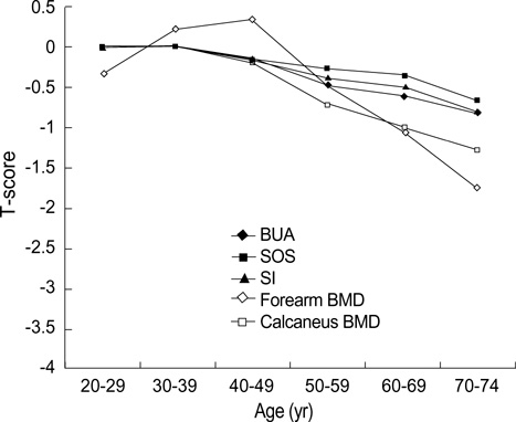
Fig. 1 Age-related decline of T-score for QUS parameters and BMD in men.
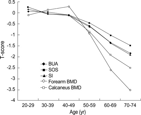
Fig. 2 Age-related decline of T-score for QUS parameters and BMD in women.
Reference
-
1. Consensus development conference. Diagnosis, prophylaxis, and treatment of osteoporosis. Am J Med. 1993. 94:646–650.2. Brandenburger GH. Clinical determination of bone quality: is ultrasound an answer? Calcif Tissue Int. 1993. 53:S151–S156.
Article3. Hans D, Dargent-Molina P, Schott AM, Sebert JL, Cormier C, Kotzki PO, Delmas PD, Pouilles JM, Breart G, Meunier PJ. Ultrasonographic heel measurements to predict hip fracture in elderly women: the EPIDOS prospective study. Lancet. 1996. 348:511–514.
Article4. Bauer DC, Gluer CC, Cauley JA, Vogt TM, Ensrud KE, Genant HK, Black DM. Study of Osteoporotic Fractures Research Group. Broadband ultrasound attenuation predicts fractures strongly and independently of densitometry in older women. A prospective study. Arch Intern Med. 1997. 157:629–634.5. Khaw KT, Reeve J, Luben R, Bingham S, Welch A, Wareham N, Oakes S, Day N. Prediction of total and hip fracture risk in men and women by quantitative ultrasound of the calcaneus: EPIC-Norfolk prospective population study. Lancet. 2004. 363:197–202.
Article6. Magkos F, Manios Y, Babaroutsi E, Sidossis LS. Contralateral differences in quantitative ultrasound of the heel: the importance of side in clinical practice. Osteoporos Int. 2005. 16:879–886.
Article7. WHO Study Group. Assessment of fracture risk and its application to screening for postmenopausal osteoporosis. Report of a WHO Study Group. World Health Organ Tech Rep Ser. 1994. 843:1–129.8. Petley GW, Cotton AM, Murrills AJ, Taylor PA, Cooper C, Cawley MI, Wilkin TJ. Reference ranges of bone mineral density for women in southern England: the impact of local data on the diagnosis of osteoporosis. Br J Radiol. 1996. 69:655–660.
Article9. Diaz Curiel M, Carrasco de la Pena JL, Honorato Perez J, Perez Cano R, Rapado A, Ruiz Martinez I. Study of bone mineral density in lumbar spine and femoral neck in a Spanish population. Multicentre Research Project on Osteoporosis. Osteoporos Int. 1997. 7:59–64.10. Looker AC, Wahner HW, Dunn WL, Calvo MS, Harris TB, Heyse SP, Johnston CC Jr, Lindsay R. Updated data on proximal femur bone mineral levels of US adults. Osteoporos Int. 1998. 8:468–489.
Article11. Chobanian AV, Bakris GL, Black HR, Cushman WC, Green LA, Izzo JL Jr, Jones DW, Materson BJ, Oparil S, Wright JT Jr, Roccella EJ. The seventh report of the joint national committee on prevention, detection, evaluation, and treatment of high blood pressure: the JNC 7 report. JAMA. 2003. 289:2560–2572.
Article12. Kim CH, Kim YI, Choi CS, Park JY, Lee MS, Lee SI, Kim GS. Prevalence and risk factors of low quantitative ultrasound values of calcaneus in Korean elderly women. Ultrasound Med Biol. 2000. 26:35–40.
Article13. Takeda N, Miyake M, Kita S, Tomomitsu T, Fukunaga M. Sex and age patterns of quantitative ultrasound densitometry of the calcaneus in normal Japanese subjects. Calcif Tissue Int. 1996. 59:84–88.
Article14. Heldan de Moura Castro C, Medeiros Pinheiro M, Lucia Szejnfeld V. Quantitative ultrasound of the calcaneus in Brazilian Caucasian women: normative data are similar to the manufacturer's normal range. Osteoporos Int. 2000. 11:923–928.15. VanderJagt DJ, Damiani LA, Goodman TM, Ujah IO, Obadofin MO, Imade GE, Shatima DR, Glew RH. Assessment of the skeletal health of healthy Nigerian men and women using quantitative ultrasound. Bone. 2004. 35:387–394.
Article16. Shin A, Choi JY, Chung HW, Park SK, Shin CS, Choi YH, Cho SI, Kim DS, Kim DI, Lee KM, Lee KH, Yoo KY, Kang D. Prevalence and risk factors of distal radius and calcaneus bone mineral density in Korean population. Osteoporos Int. 2004. 15:639–644.
Article17. Gallagher JC, Goldgar D, Moy A. Total bone calcium in normal women: effect of age and menopause status. J Bone Miner Res. 1987. 2:491–496.
Article18. Nuti R, Righi G, Martini G, Turchetti V, Lepore C, Caniggia A. Methods and clinical applications of total body absorptiometry. J Nucl Med Allied Sci. 1987. 31:213–221.
Article19. Kaneko M, Miyake T, Yokoyama E, Harano S, Toki T, Komine Y, Nozaki N, Nozaki S, Takeda N, Miyake M, Fukunaga M. Standard radial bone mineral density and physical factors in ordinary Japanese women. J Bone Miner Metab. 2000. 18:31–35.
Article20. Berntsen GK, Fonnebo V, Tollan A, Sogaard AJ, Magnus JH. Forearm bone mineral density by age in 7,620 men and women: the Tromso study, a population-based study. Am J Epidemiol. 2001. 153:465–473.21. Forsmo S, Langhammer A, Forsen L, Schei B. Forearm bone mineral density in an unselected population of 2,779 men and women: The HUNT Study, Norway. Osteoporos Int. 2005. 16:562–567.22. Frost ML, Blake GM, Fogelman I. Quantitative ultrasound and bone mineral density are equally strongly associated with risk factors for osteoporosis. J Bone Miner Res. 2001. 16:406–416.
Article23. Knapp KM, Blake GM, Spector TD, Fogelman I. Can the WHO definition of osteoporosis be applied to multi-site axial transmission quantitative ultrasound? Osteoporos Int. 2004. 15:367–374.
Article24. Kanis JA, Gluer CC. An update on the diagnosis and assessment of osteoporosis with densitometry. Committee of scientific advisors, international osteoporosis foundation. Osteoporos Int. 2000. 11:192–202.25. Gregg EW, Kriska AM, Salamone LM, Roberts MM, Anderson SJ, Ferrell RE, Kuller LH, Cauley JA. The epidemiology of quantitative ultrasound: a review of the relationships with bone mass, osteoporosis and fracture risk. Osteoporos Int. 1997. 7:89–99.
Article26. Waud CE, Lew R, Baran DT. The relationship between ultrasound and densitometric measurements of bone mass at the calcaneus in women. Calcif Tissue Int. 1992. 51:415–418.
Article27. Salamone LM, Krall EA, Harris S, Dawson-Hughes B. Comparison of broadband ultrasound attenuation to single X-ray absorptiometry measurements at the calcaneus in postmenopausal women. Calcif Tissue Int. 1994. 54:87–90.
Article28. Ross P, Huang C, Davis J, Imose K, Yates J, Vogel J, Wasnich R. Predicting vertebral deformity using bone densitometry at various skeletal sites and calcaneus ultrasound. Bone. 1995. 16:325–332.
Article
- Full Text Links
-
- Actions
-
Cited
- CITED
-
- Close
- Share
-
- Similar articles
-
- Measurement of bone mineral density in Korean newborns by dual photon absorptiometry
- Reliability of Singh's index Checked by the Dual Photon X-ray Absorptiometry(LUNAR D.P.X)
- Measurement and Interpretation of Dual-Energy X-ray Absorptiometry Bone Density Measurements
- Pediatric dual-energy X-ray absorptiometry: interpretation and clinical and research application
- Cross-calibration of Bone Mineral Density between Two Different Dual X-ray Absorptiometry Systems: Hologic QDR 4500-A and Lunar EXPERT-XL
