A Case of Poikiloderma Vasculare Atrophicans
- Affiliations
-
- 1Department of Dermatology, Chonnam National University Medical School, Gwangju, Korea. sjyun@chonnam.ac.kr
- KMID: 2156750
- DOI: http://doi.org/10.5021/ad.2011.23.S1.S48
Abstract
- Poikiloderma vasculare atrophicans (PVA) is a rare variant of mycosis fungoides, and is characterized by generalized hyperkeratotic scaly papules in net-like, retiform, or zebra-like patterns. A 59-year-old Korean woman presented with asymptomatic, erythematous-to-violaceous, reticulated confluent papules on the trunk and extremities. Skin lesions were initially limited to both thighs 25 years ago, and then spread slowly over her body. Histopathological examination showed band-like inflammatory infiltrations and epidermotropism consisting of mostly CD8+ lymphocytes. Based on the clinical manifestations and histological findings, the diagnosis of PVA was made. We herein report on a case of PVA, which featured a long-benign course without progression into the tumor stage over a period of 30 years.
Figure
-
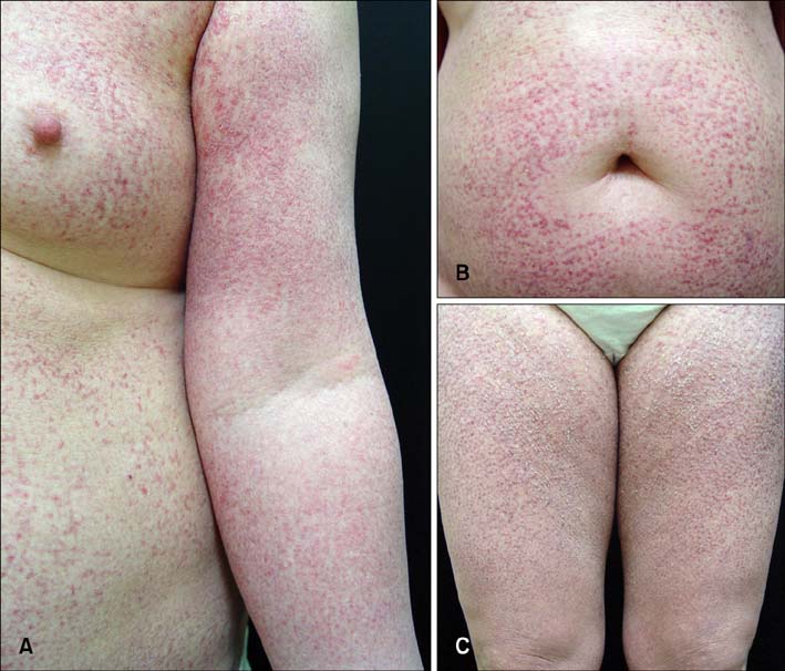
Fig. 1 Erythematous hyperkeratotic confluent papules with a reticulated or net-like pattern on her trunk and upper extremities (A), abdomen (B), and both thighs (C).
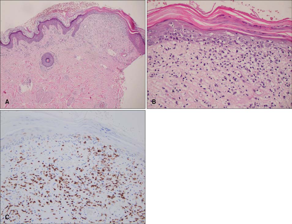
Fig. 2 Skin biopsy specimen from the thigh in 2006 shows compact hyperkeratosis, thinned epidermis, and band-like inflammatory cell infiltrations of mostly lymphocytes with dilated capillaries in the upper dermis (A, H&E; ×40). On high power view, focal parakeratosis, epidermotropism of atypical lymphocytes in the epidermis, and abnormal, wiry patterned collagen bundles in the upper dermis (B, H&E; ×200), and a CD8-positive reaction of infiltrative lymphocytes in immunochemical study (C, CD8; ×200) are shown.
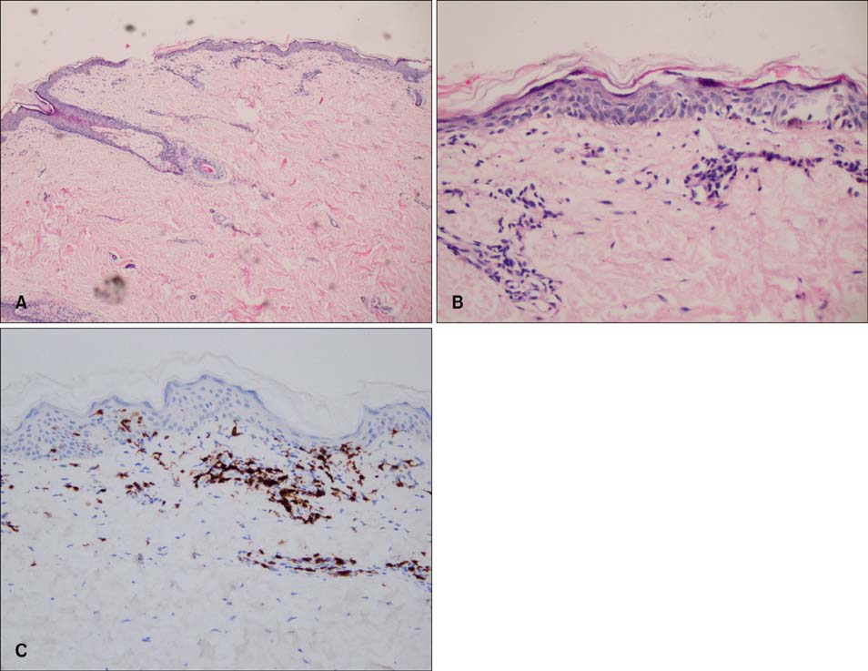
Fig. 3 Skin biopsy specimen from the thigh 25 years ago shows perivascular inflammatory cell infiltrations of mostly lymphocytes in the upper dermis (A, H&E; ×40), epidermotropism of atypical lymphocytes in the epidermis (B, H&E; ×200), and a CD8- positive reaction of infiltrative lymphocytes in immunochemical staining (C, CD8; ×200).
Cited by 1 articles
-
Poikiloderma Vasculare Atrophicans Showing Features of Ashy Dermatosis in the Beginning
Jiehyun Jeon, Joo Ha Kim, Jae Woo Ahn, Hae Jun Song
Ann Dermatol. 2015;27(2):197-200. doi: 10.5021/ad.2015.27.2.197.
Reference
-
1. Brecher A. Mycosis fungoides. Dermatol Online J. 2003. 9:23.
Article2. Kikuchi A, Naka W, Nishikawa T. Cutaneous T-cell lymphoma arising from parakeratosis variegata: long-term observation with monitoring of T-cell receptor gene rearrangements. Dermatology. 1995. 190:124–127.
Article3. Kazakov DV, Burg G, Kempf W. Clinicopathological spectrum of mycosis fungoides. J Eur Acad Dermatol Venereol. 2004. 18:397–415.
Article4. Assaf C, Sterry W. Wolff K, Goldsmith LA, Katz SI, Gilchrest BA, Paller AS, Leffell DJ, editors. Cutaneous lymphoma. Fitzpatrick's dermatology in general medicine. 2008. 7th ed. New York, USA: McGraw-Hill;2154–2157.5. Christine J. Elder DE, Elenitsas R, Johnson BL, Murphy GF, editors. Connective tissue disease. Lever's histopahology of the skin. 2005. 9th ed. Philadelphia, USA: Lippincott Williams & Wilkins;308–309.6. McKee PH, Calonje E, Granter SR. Pathology of the skin with clinical correlations. 2005. 3rd ed. Philadelphia, USA: Elsevier Mosby;1763–1764.7. Kreuter A, Hoffmamm K, Altmeyer P. A case of poikiloderma vasculare atrophicans, a rare variant of cutaneous T-cell lymphoma, responding to extracorporeal photopheresis. J Am Acad Dermatol. 2005. 52:706–708.
Article8. Lee JB, Myung KB, Kim JH. Parapsoriasis variegata: report of a case. Korean J Dermatol. 1979. 17:367–371.9. Jang HC, Ahn BJ, Kim SW, Kim DS. A case of poikilodermatous mycosis fungoides. Korean J Dermatol. 1999. 37:1515–1517.10. Cho KH, Lee HS. A clinicopathological study of early mycosis fungoides. Korean J Dermatol. 1999. 37:838–845.11. Lu D, Patel KA, Duvic M, Jones D. Clinical and pathological spectrum of CD8-positive cutaneous T-cell lymphomas. J Cutan Pathol. 2002. 29:465–472.
Article12. Berti E, Tomasini D, Vermeer MH, Meijer CJ, Alessi E, Willemze R. Primary cutaneous CD8-positive epidermotropic cytotoxic T cell lymphomas. A distinct clinicopathological entity with an aggressive clinical behavior. Am J Pathol. 1999. 155:483–492.
Article13. Agnarsson BA, Vonderheid EC, Kadin ME. Cutaneous T cell lymphoma with suppressor/cytotoxic (CD8) phenotype: identification of rapidly progressive and chronic subtypes. J Am Acad Dermatol. 1990. 22:569–577.
Article14. Santucci M, Pimpinelli N, Massi D, Kadin ME, Meijer CJ, Müller-Hermelink HK, et al. Cytotoxic/natural killer cell cutaneous lymphomas. Report of EORTC Cutaneous Lymphoma Task Force Workshop. Cancer. 2003. 97:610–627.15. Nikolaou VA, Papadavid E, Katsambas A, Stratigos AJ, Marinos L, Anagnostou D, et al. Clinical characteristics and course of CD8+ cytotoxic variant of mycosis fungoides: a case series of seven patients. Br J Dermatol. 2009. 161:826–830.
Article16. van Krieken JH, Langerak AW, Macintyre EA, Kneba M, Hodges E, Sanz RG, et al. Improved reliability of lymphoma diagnostics via PCR-based clonality testing: report of the BIOMED-2 Concerted Action BHM4-CT98-3936. Leukemia. 2007. 21:201–206.
Article17. Thurber SE, Zhang B, Kim YH, Schrijver I, Zehnder J, Kohler S. T-cell clonality analysis in biopsy specimens from two different skin sites shows high specificity in the diagnosis of patients with suggested mycosis fungoides. J Am Acad Dermatol. 2007. 57:782–790.
Article18. Nashan D, Faulhaber D, Ständer S, Luger TA, Stadler R. Mycosis fungoides: a dermatological masquerader. Br J Dermatol. 2007. 156:1–10.
Article
