A Case of an Unusual Eccrine Poroma on the Left Forearm Area
- Affiliations
-
- 1Department of Dermatology, School of Medicine, Keimyung University, Daegu, Korea. janylove99@dsmc.or.kr
- KMID: 2156676
- DOI: http://doi.org/10.5021/ad.2011.23.2.250
Abstract
- A 40-year-old woman presented with an asymptomatic red to brown colored walnut-sized, dome shaped, hemorrhagic, crusted nodule on the left forearm. There was no previous history of trauma to the area. The first impression of this case was a vascular tumor or malignant lesion due to the large size and bleeding tendency. However, the final diagnosis, according to histologic and immunostaining methods, was a benign eccrine poroma that occurred on the left forearm, which is an unusual area for such a lesion. The tumor was excised and no recurrence was noted when she was examined 24 months later.
Keyword
Figure
-

Fig. 1 (A) A solitary 2.0×2.0 cm sized red to brown-colored mass on the left forearm. (B) The mass seemed to be pedunculated and it had a rough surface that showed hemorrhagic crusts and erosions.
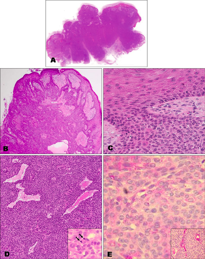
Fig. 2 (A) A single pedunculated large tumor mass and well-circumscribed tumor nests within the epidermis and these nests were located at the periphery of the mass (H&E, 1:1 direct view). (B) This scanning view showed the interconnected epithelial downward growth with multiple foci of attachment to the epidermis (H&E, ×40). (C) The tumor cells were small and they showed a uniform cuboidal appearance with basophilic, round nuclei and the tumor cells were connected by intercellular bridges (H&E, ×400). (D) There were conspicuous intracytoplasmic lumina (H&E, ×100). The tumor cells showed slightly increased mitotic figures (inset). (E) The tumor cells contained periodic acid-Schiff (PAS)-positive materials (H&E, ×200) and d-PAS positive materials (inset).

Fig. 3 (A) The ductal lining stained positive for carcinoembryogenic antigen (H&E, ×100). The tumor cells stained positive for epithelial membrane antigen (B: H&E, ×100), negative for S-100 (C: H&E, ×100), and less than 10% of the tumor cells were positive for Ki-67 (D: H&E, ×200) and p53 (E: H&E, ×200).
Reference
-
1. Goldman P, Pinkus H, Rogin JR. Eccrine poroma; tumors exhibiting features of the epidermal sweat duct unit. AMA Arch Derm. 1956. 74:511–521.2. LoBuono P, Kahn R, Kornblee LV. Eccrine poroma of the forehead. Mt Sinai J Med. 1977. 44:527–529.3. Jin WW, Jung JG, Ro KW, Shim SD, Kim MH, Cinn YW. A case of eccrine poroma on the paranasal area. Ann Dermatol. 2006. 18:73–76.
Article4. Park SJ, Roh BH, Lee JS, Cho MK, Whang KU. A case of eccrine poroma on the scalp. Korean J Dermatol. 2006. 44:633–635.5. Johnson RC, Rosenmeier GJ, Keeling JH 3rd. A painful step. Eccrine poroma. Arch Dermatol. 1992. 128:1530.
Article6. Kircik L, Armus S, Kipping H, Pincus SH. Eccrine poroma in an unusual location. Cutis. 1994. 54:183–184.7. Pylyser K, De Wolf-Peeters C, Marien K. The histology of eccrine poromas: a study of 14 cases. Dermatologica. 1983. 167:243–249.
Article8. Harvell JD, Kerschmann RL, LeBoit PE. Eccrine or apocrine poroma? Six poromas with divergent adnexal differentiation. Am J Dermatopathol. 1996. 18:1–9.9. Robson A, Greene J, Ansari N, Kim B, Seed PT, McKee PH, et al. Eccrine porocarcinoma (malignant eccrine poroma): a clinicopathologic study of 69 cases. Am J Surg Pathol. 2001. 25:710–720.10. Shaw M, McKee PH, Lowe D, Black MM. Malignant eccrine poroma: a study of twenty-seven cases. Br J Dermatol. 1982. 107:675–680.
Article11. Pinkus H, Mehregan AH. Epidermotrophic eccrine carcinoma. A case combining features of eccrine poroma and Paget's dermatosis. Arch Dermatol. 1963. 88:597–606.12. Witkowski JA, Parish LC, Griffith CQ. Solitary eccrine poroma. Int J Dermatol. 1979. 18:307–308.
Article
