Ann Dermatol.
2009 Nov;21(4):389-392. 10.5021/ad.2009.21.4.389.
Ecthyma Gangrenosum: A Rare Cutaneous Manifestation Caused by Stenotrophomonas maltophilia in a Leukemic Patient
- Affiliations
-
- 1Department of Dermatology, Gachon University of Medicine and Science, Gil Medical Center, Incheon, Korea. jyroh1@gilhospital.com
- KMID: 2156502
- DOI: http://doi.org/10.5021/ad.2009.21.4.389
Abstract
- Ecthyma gangrenosum (EG) is a well-recognized cutaneous infection that most commonly affects immunocompromised patients. It typically occurs on the extremities, or in gluteal and perineal regions. Although Pseudomonas aeruginosa is the most well-known pathogen causing EG, other organisms have been reported to cause EG. Herein we report a rare case of ecthyma gangrenosum presenting as aggressive necrotic skin lesions in perioral and infraorbital areas in a 47-year-old patient with acute myelocytic leukemia after allogeneic bone marrow transplantation. It was caused by Stenotrophomonas maltophilia, which is an aerobic, gram-negative pathogen that has been associated only rarely with cutaneous disease. Blood culture and tissue culture were positive for S. maltophilia. Histological examination revealed numerous tiny bacilli in the dermis and perivascular area. Early recognition of skin lesions caused by S. maltophilia is important to decrease associated mortality in immunosuppressed patients.
MeSH Terms
Figure
-

Fig. 1 Severe edematous erythematous necrotic plaques with perioral bullae formation.
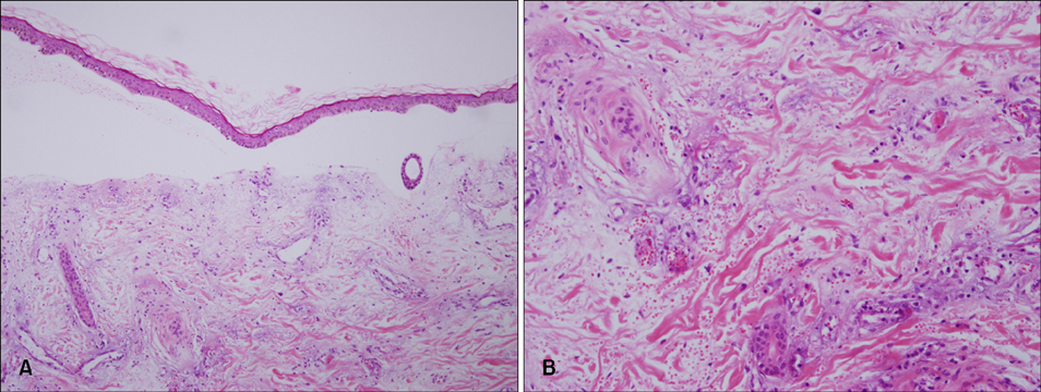
Fig. 2 (A) Skin biopsy specimen showing subepidermal blister with hemorrhage and dermal edema with vascular congestion (H&E, ×100). (B) Perivascular cuffing with basophilic deposits accompanied by a mixed inflammatory infiltrate of lymphocytes and histiocytes in dermis. Intravascular thrombosis was present with endothelial swelling (H&E, ×200).
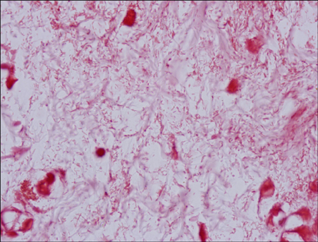
Fig. 3 Numerous gram-negative bacilli surround papillary dermal vessels and infiltrate the tissue (Gram stain, ×1,000).
Reference
-
1. James WD, Berger TG, Elston DM. Andrews' diseases of the skin: clinical dermatology. 2006. 10th ed. Philadelphia: Saunders Elsevier;271–272.2. McKee PH, Calonje E, Granter SR. Pathology of the skin: with clinical correlations. 2005. 3rd ed. Philadelphia: Elsevier Mosby;869–871.3. Downey DM, O'Bryan MC, Burdette SD, Michael JR, Saxe JM. Ecthyma gangrenosum in a patient with toxic epidermal necrolysis. J Burn Care Res. 2007. 28:198–202.
Article4. Pandit AM, Siddaramappa B, Choudhary SV, Manjunathswamy BS. Ecthyma gangrenosum in a new born child. Indian J Dermatol Venereol Leprol. 2003. 69:52–53.5. Reich HL, Williams Fadeyi D, Naik NS, Honig PJ, Yan AC. Nonpseudomonal ecthyma gangrenosum. J Am Acad Dermatol. 2004. 50:5 Suppl. S114–S117.
Article6. Song WK, Kim YC, Park HJ, Cinn YW. Ecthyma gangrenosum without bacteraemia in a leukaemic patient. Clin Exp Dermatol. 2001. 26:395–397.
Article7. Lee GY, Choi YW, Choi HY, Myung KB. A case of onychia and paronychia by Sternotrophomonas maltophilia. Korean J Dermatol. 2001. 39:89–90.8. Teo WY, Chan MY, Lam CM, Chong CY. Skin manifestation of Stenotrophomonas maltophilia infection--a case report and review article. Ann Acad Med Singapore. 2006. 35:897–900.9. Sakhnini E, Weissmann A, Oren I. Fulminant Stenotrophomonas maltophilia soft tissue infection in immunocompromised patients: an outbreak transmitted via tap water. Am J Med Sci. 2002. 323:269–272.
Article10. Moser C, Jonsson V, Thomsen K, Albrectsen J, Hansen MM, Prag J. Subcutaneous lesions and bacteraemia due to Stenotrophomonas maltophilia in three leukaemic patients with neutropenia. Br J Dermatol. 1997. 136:949–952.
Article11. Solowski NL, Yao FB, Agarwal A, Nagorsky M. Ecthyma gangrenosum: a rare cutaneous manifestation of a potentially fatal disease. Ann Otol Rhinol Laryngol. 2004. 113:462–464.
Article12. Zomorrodi A, Wald ER. Ecthyma gangrenosum: considerations in a previously healthy child. Pediatr Infect Dis J. 2002. 21:1161–1164.
Article13. Singh TN, Devi KM, Devi KS. Ecthyma gangrenosum: a rare cutaneous manifestation caused by Pseudomonas aeruginosa without bacteraemia in a leukaemic patient--a case report. Indian J Med Microbiol. 2005. 23:262–263.
Article14. Boisseau AM, Sarlangue J, Perel Y, Hehunstre JP, Taieb A, Maleville J. Perineal ecthyma gangrenosum in infancy and early childhood: septicemic and nonsepticemic forms. J Am Acad Dermatol. 1992. 27:415–418.
Article15. Rodot S, Lacour JP, van Elslande L, Castanet J, Desruelles F, Ortonne JP. Ecthyma gangrenosum caused by Klebsiella pneumoniae. Int J Dermatol. 1995. 34:216–217.
Article16. Smeets JG, Lowe SH, Veraart JC. Cutaneous infections with Stenotrophomonas maltophilia in patients using immunosuppressive medication. J Eur Acad Dermatol Venereol. 2007. 21:1298–1300.17. Dignani MC, Grazziutti M, Anaissie E. Stenotrophomonas maltophilia infections. Semin Respir Crit Care Med. 2003. 24:89–98.18. Jang TN, Wang FD, Wang LS, Liu CY, Liu IM. Xanthomonas maltophilia bacteremia: an analysis of 32 cases. J Formos Med Assoc. 1992. 91:1170–1176.19. Betriu C, Sanchez A, Palau ML, Gomez M, Picazo JJ. Antibiotic resistance surveillance of Stenotrophomonas maltophilia, 1993-1999. J Antimicrob Chemother. 2001. 48:152–154.
Article20. del Toro MD, Rodriguez-Bano J, Herrero M, Rivero A, Garcia-Ordonez MA, Corzo J, et al. Clinical epidemiology of Stenotrophomonas maltophilia colonization and infection: a multicenter study. Medicine (Baltimore). 2002. 81:228–239.
- Full Text Links
-
- Actions
-
Cited
- CITED
-
- Close
- Share
-
- Similar articles
-
- Clinical Examination of Stenotrophomonas maltophilia Infection in Patient with Pyoderma Gangrenosum
- Localized Cutaneous Infection due to Stenotrophomonas maltophilia in Immunocompetent Patient
- A Case of Corneal Ulcer Caused by Combined Infection of Stenotrophomonas Maltophilia and Aspergillus Fumigatus
- Stenotrophomonas maltophilia Periprosthetic Joint Infection after Hip Revision Arthroplasty
- A Case of Metastatic Cellulitis Caused by Stenotrophomonas maltophilia
