Arthroscopic Resection of Osteochondroma of Hip Joint Associated with Internal Snapping: A Case Report
- Affiliations
-
- 1Department of Orthopedic Surgery, Busan Bumin Hospital, Busan, Korea.
- 2Department of Orthopedic Surgery, Chungnam National University School of Medicine, Daejeon, Korea.
- 3Department of Orthopedic Surgery, Seoul Bumin Hospital, Seoul, Korea. mskps@naver.com
- KMID: 2156019
- DOI: http://doi.org/10.5371/hp.2015.27.1.43
Abstract
- A 16-year old male patient visited the hospital complaining of inguinal pain and internal snapping of right hip joint. In physical examination, the patient was presumed to be diagnosed femoroacetabular impingement (FAI) and acetabular labral tear. In radiologic evaluation, FAI and acetabular labral tear were identified and bony tumor associated with internal snapping was found on the posteromedial portion of the femoral neck. Despite of conservative treatment, there was no symptomatic improvement. So arthroscopic labral repair, osteoplasty and resection of bony tumor were performed. The tumor was pathologically diagnosed as osteochondroma through biopsy and all symptoms improved after surgery. There was no recurrence, complication or abnormal finding during 1 year follow up. Osteochondroma located at posteromedial portion of femoral neck can be a cause of internal snapping hip and although technical demands are challenging, arthroscopic resection can be a good treatment option.
Keyword
MeSH Terms
Figure
-
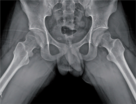
Fig. 1 Frog-leg lateral view. The bony lesion was found at posteromedial portion of right femoral neck.
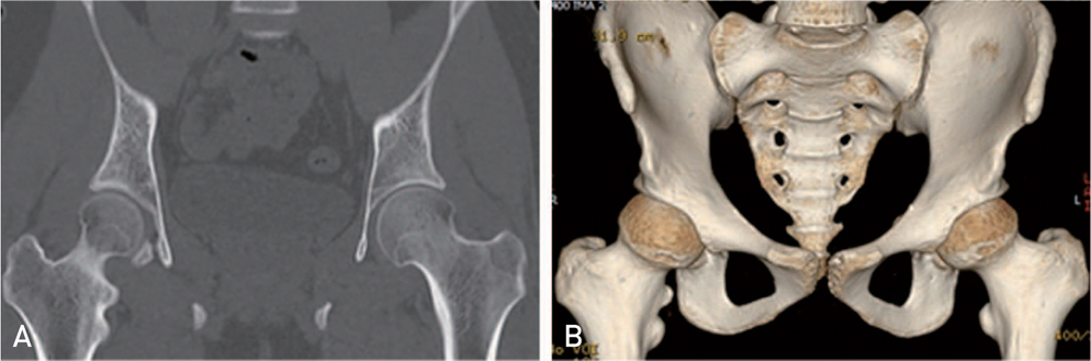
Fig. 2 (A) Preoperative computed tomography (CT). Bony protuberance was located just above lesser trochanter. (B) Preoperative CT. Bony protuberance was located just above lesser trochanter.
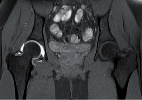
Fig. 3 Intraarticular bony lesion was suggestive of osteochondroma with cartilaginous cap in magnetic resonance arthrography image.

Fig. 4 (A) Arthroscopic finding of acetabular labral tear (arrow) at anterosuperior portion of acetabulum. Anterolateral and anterior portal were used as viewing and working portal, each. (B) Acetabuloplasty using arthroscopic spherical burr (Linvatec, Largo, FL, USA) for correction of pincer femoroacetabular impingement. (C) Acetabular labral repair with two bioabsorbable suture anchors (Bioraptor; Smith & Nephew, Andover, MA, USA).

Fig. 5 Arthroscopic finding of osteochondroma with cartilaginous cap (arrow) at posteromedial portion of femoral neck. FN: femoral neck, OC: osteochondroma.
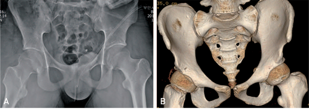
Fig. 6 (A) Postoperative simple radiography. The bony mass was removed successfully. (B) Postoperative 3 dimensional computed tomography image. Complete resection of osteochondroma was identified and degree of osteoplasty was evaluated postoperatively.

Fig. 7 The cartilagious cap was identified in biopsy and the cap has a smooth round surface of a pathologic finding of osteochondroma (hematoxylin and eosin stain, ×40).
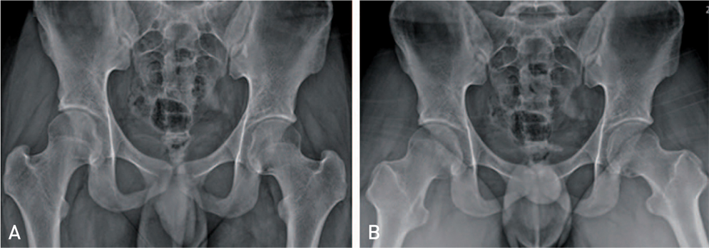
Fig. 8 (A) Hip anteroposterior simple radiopraphy at postoperative 1 year. Posteromedial cortex of the femoral neck was reformed and any bony recurrence was not shown. (B) Hip frog leg lateral radiography at postoperative 1 year.
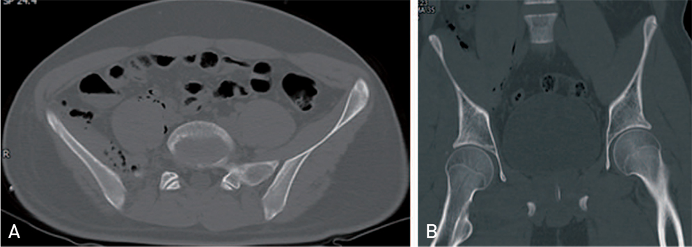
Fig. 9 (A) In postoperative axial image, leakage of arthroscopic fluid and air bubbles were shown around iliopsoas muscle. (B) In coronal computed tomography image, air bubbles were found along iliopsoas muscle.
Reference
-
1. Kanauchi T, Suganuma J, Kawasaki T, et al. Fracture of an osteochondroma of the femoral neck caused by impingement against the ischium. Orthopedics. 2012; 35:e1438–e1441.
Article2. Yu K, Meehan JP, Fritz A, Jamali AA. Osteochondroma of the femoral neck: a rare cause of sciatic nerve compression. Orthopedics. 2010; 33:DOI: 10.3928/01477447-20100625-26.
Article3. Inoue S, Noguchi Y, Mae T, Rikimaru S, Hotokezaka S. An external snapping hip caused by osteochondroma of the proximal femur. Mod Rheumatol. 2005; 15:432–434.
Article4. Hussain W, Avedian R, Terry M, Peabody T. Solitary osteochondroma of the proximal femur and femoral acetabular impingement. Orthopedics. 2010; 33:51.
Article5. Siebenrock KA, Ganz R. Osteochondroma of the femoral neck. Clin Orthop Relat Res. 2002; 394:211–218.
Article6. Carpintero P, León F, Zafra M, Montero M, Berral FJ. Fractures of osteochondroma during physical exercise. Am J Sports Med. 2003; 31:1003–1006.
Article7. Dienst M, Gödde S, Seil R, Hammer D, Kohn D. Hip arthroscopy without traction: In vivo anatomy of the peripheral hip joint cavity. Arthroscopy. 2001; 17:924–931.8. Feeley BT, Kelly BT. Arthroscopic management of an intraarticular osteochondroma of the hip. Orthop Rev (Pavia). 2009; 1:e2.
Article9. Lee JB, Kang C, Lee CH, Kim PS, Hwang DS. Arthroscopic treatment of synovial chondromatosis of the hip. Am J Sports Med. 2012; 40:1412–1418.
Article
- Full Text Links
-
- Actions
-
Cited
- CITED
-
- Close
- Share
-
- Similar articles
-
- Osteochondroma Arising from Anterior Inferior Iliac Spine as a Cause of Snapping Hip
- Painful Snapping Shoulder Complicating Soft Tissue Pseudotumor Secondary to Rib Osteochondroma: A Case Report
- Arthroscopic Excision of Intra-articular Osteochondroma of the Elbow: A Case Report
- Snapping Knee caused by the Gracilis and Semitendinosus tendon
- Arthroscopic Treatment of Symptomatic Internal Snapping Hip with Combined Pathologies
