Changes in Tip-Apex Distance by Position and Film Distance Measured by Picture Archiving and Communication System (PACS)
- Affiliations
-
- 1Department of Orthopaedic Surgery, Dong-A University College of Medicine, Busan, Korea. hyeonjun@dau.ac.kr
- 2Department of Diagnostic Radiology, Dong-A University College of Medicine, Busan, Korea.
- KMID: 2156018
- DOI: http://doi.org/10.5371/hp.2015.27.1.36
Abstract
- PURPOSE
The tip-apex distance (TAD) is used to predict the clinical outcome of intertrochanteric fracture fixation. We aimed to measure the changes in TAD by position and film distance using Picture Archiving and Communication System (PACS).
MATERIALS AND METHODS
We used a femur replica with a 10degrees femoral neck anteversion and a 130degrees neck shaft angle. Proximal femoral nail antirotation nail and a helical blade were inserted into the replica. Radiographs were taken at the neutral position and after applying 10degrees, 20degrees, 30degrees, 40degrees internal/external rotation, 10degrees abduction, and 10degrees and 40degrees adduction to the mechanical axis. Radiographs were taken at the replica-film distance of 10 cm and 20 cm under the same conditions, mimicking the differences in Focus-film distance (FFD), which reflect the patient's contour in clinical settings. A radiologist and an orthopedic surgeon measured the TAD twice using PACS. The average error was 2 mm (4.5%) and the standard error was +/-3.04. TADs in the neutral position constituted the standard values to measure the relative errors.
RESULTS
TADs increased with an increase in the external rotation and abduction of the replica. TADs decreased with an increase in the internal rotation and adduction of the replica. For comparable measurements, relative errors were higher at FFDs of 20 cm compared to FFDs of 10 cm.
CONCLUSION
Since the femur is internally rotated and adducted for reduction, orthopedic surgeons would assess the lag screw to be closer to the apex of femur on intraoperative radiographs. To have a correct measurement of the TAD after fixation of intertrochanteric fractures, radiographs should be taken in neutral position and measurement errors should be considered based on the patient's size.
Figure
-
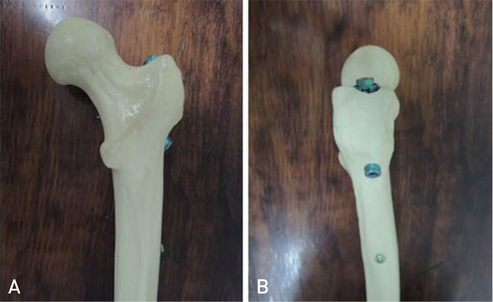
Fig. 1 Proximal femoral nail antirotation nail and a helical blade inserted into the femur replica. (A) Anteroposterior and (B) lateral views.
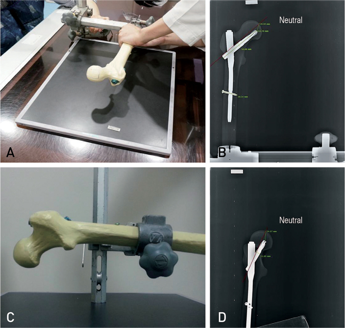
Fig. 2 (A) Radiograph is taken after applying internal/external rotation to the mechanical axis. (B) Anteroposterior radiograph in neutral position. (C) Radiograph is taken after applying abduction/adduction to the mechanical axis. (D) Lateral radiograph in 15° external rotation.
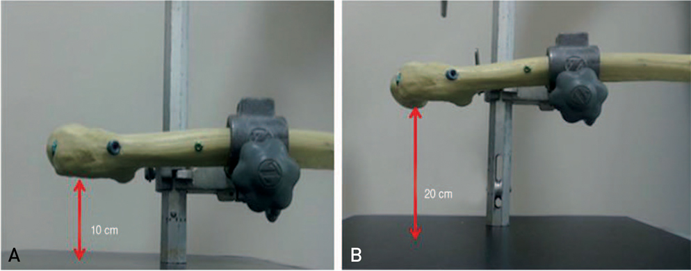
Fig. 3 Radiographs are taken at replica-film distances of 10 cm and 20 cm under same conditions.
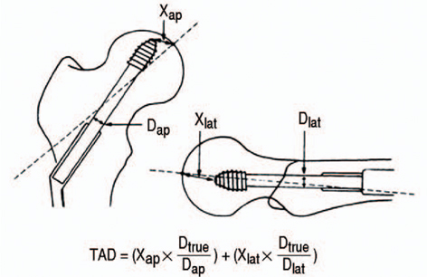
Fig. 4 Tip-apex distance (TAD) is the summation of the distance, in millimeters, from the tip of the lag screw to the apex of the femoral head on the anteroposterior (ap) and lateral (lat) radiographs.
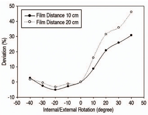
Fig. 5 Tip-apex distance (TAD) on the anteroposterior view.
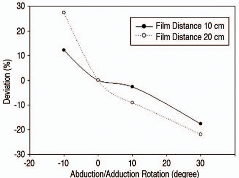
Fig. 6 Tip-apex distance (TAD) on the lateral view.
Reference
-
1. Kelly RF. Fracutres of the proximal part of the femur. J Bone Joint Surg Am. 1994; 76:924–950.2. Park YS, Park YK, Choi HJ. Result of bilateral total hip arthroplasty: one stage versus two stage procedure. J Korean Hip Soc. 1998; 10:211–215.3. Suh JT, Chung WB, Yoo CI. A clinical study of trochanteric fractures of the femur in the elderly over 70 years in the age. J Korean Soc Fract. 1994; 7:293–301.
Article4. Boriani S, Bettelli G, Zmerly H, et al. Results of the multicentric Italian experience on the Gamma nail: a report on 648 cases. Orthopedics. 1991; 14:1307–1314.
Article5. Davis J, Harris MB, Duval M, D'Ambrosia R. Pertrochanteric fractures treated with the Gamma nail: technique and report of early results. Orthopedics. 1991; 14:939–942.
Article6. Geller JA, Saifi C, Morrison TA, Macaulay W. Tip-apex distance of intramedullary devices as a predictor of cut-out failure in the treatment of peritrochanteric elderly hip fractures. Int Orthop. 2010; 34:719–722.
Article7. Forte ML, Virnig BA, Kane RL, et al. Geographic variation in device use for intertrochanteric hip fractures. J Bone Joint Surg Am. 2008; 90:691–699.
Article8. Lee KJ, Min BW, Kim SG, Song KS, Bae KC, Cho CH. Result of treating sensile osteoporotic peritrochanteric fracture with proximal femoral nail antirotation (PNFA). J Korean Hip Soc. 2009; 21:162–168.
Article9. Yoo JH, Park JS, Noh KC, et al. The results of proximal femoral nail antirotation: a comparative study with proximal femoral nail. J Korean Hip Soc. 2008; 20:286–292.
Article10. Hong KD, Sim JC, Ha SS, Kim TH, Choi YH, Kim JH. Operative treatment with Gamma 3 nail in femur intertrochanteric fracture. J Korean Fract Soc. 2011; 24:7–15.
Article11. Kawaguchi S, Sawada K, Nabeta Y. Cutting-out of the lag screw after internal fixation with the Asiatic gamma nail. Injury. 1998; 29:47–53.
Article12. Nishiura T, Nozawa M, Morio H. The new technique of precise insertion of lag screw in an operative treatment of trochanteric femoral fractures with a short intramedullary nail. Injury. 2009; 40:1077–1083.
Article13. Baumgaertner MR, Curtin SL, Lindskog DM, Keggi JM. The value of the tip-apex distance in predicting failure of fixation of peritrochanteric fractures of the hip. J Bone Joint Surg Am. 1995; 77:1058–1064.
Article14. Baumgaertner MR, Solberg BD. Awareness of tip-apex distance reduces failure of fixation of trochanteric fractures of the hip. J Bone Joint Surg Br. 1997; 79:969–971.
Article15. White SP, Shardlow DL. Effect of introduction of digital radiographic techniques on pre-operative templating in orthopaedic practice. Ann R Coll Surg Engl. 2005; 87:53–54.
Article16. Eklund K, Jonsson K, Lindblom G, et al. Are digital images good enough? A comparative study of conventional film-screen vs digital radiographs on printed images of total hip replacement. Eur Radiol. 2004; 14:865–869.
Article17. Cleveland M, Bosworth DM, Thompson FR, Wilson HJ Jr, Ishizuka T. A ten-year analysis of intertrochanteric fractures of the femur. J Bone Joint Surg Am. 1959; 41-A:1399–1408.
Article18. Nikoloski AN, Osbrough AL, Yates PJ. Should the tipapex distance (TAD) rule be modified for the proximal femoral nail antirotation (PFNA)? A retrospective study. J Orthop Surg Res. 2013; 8:35.
Article19. De Bruijn K, den Hartog D, Tuinebreijer W, Roukema G. Reliability of predictors for screw cutout in intertrochanteric hip fractures. J Bone Joint Surg Am. 2012; 94:1266–1272.
Article20. Pervez H, Parker MJ, Vowler S. Prediction of fixation failure after sliding hip screw fixation. Injury. 2004; 35:994–998.
Article21. Kim SL, Kim YM, Choi IH, Seong SC, Chung CY, Kim DH. Anthropological studies on the proximal femur of the Korean. J Korean Orthop Assoc. 1986; 21:767–780.
Article
- Full Text Links
-
- Actions
-
Cited
- CITED
-
- Close
- Share
-
- Similar articles
-
- The Value of the Tip - Apex Distance in Predicting Failure of Fixation of Intertrochanteric Fractures of the Hip
- The effect of the introduction of Picture Archiving and Communication System on interpretation rate of radiologic examinations
- Usefulness of Tip-to-Carina Distance as a Marker for Detecting Right Atrium Positioning of a Central Venous Catheter on Chest X-Ray
- Current status and installation standard of dental PACS
- Dental PACS development in Korea
