A tiny bleb at Junctional Dilatation of the Posterior Communicating Artery as a Predisposing Factor for Development of a De Novo Aneurysm
- Affiliations
-
- 1Department of Radiology, Asan Medical Center, Ulsan University School of Medicine, Seoul, Korea. dcsuh@amc.seoul.kr
- 2Department of Neurosurgery, Asan Medical Center, Ulsan University School of Medicine, Seoul, Korea.
- KMID: 2155722
- DOI: http://doi.org/10.5469/neuroint.2016.11.1.59
Abstract
- Formation of de novo aneurysm from a junctional dilatation at the origin site of the posterior communicating artery (PcomA) has been rarely reported. In this case report, three females in sixth decades of age developed a de novo aneurysm from the junctional dilatation of the PComA with a tiny bleb-like lesion over 5 years after initial presentation.
Figure
-
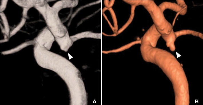
Fig. 1 A 53-year-old female (Case 1). (A) Initial 3-D rotational angiogram shows a bilobulated bleb (arrowhead) at junctional dilatation of the posterior communicating artery (PcomA). (B) Follow-up 3-D rotational angiogram obtained 5 years later shows de novo aneurysm formation at the site of the previous bleb (arrowhead).
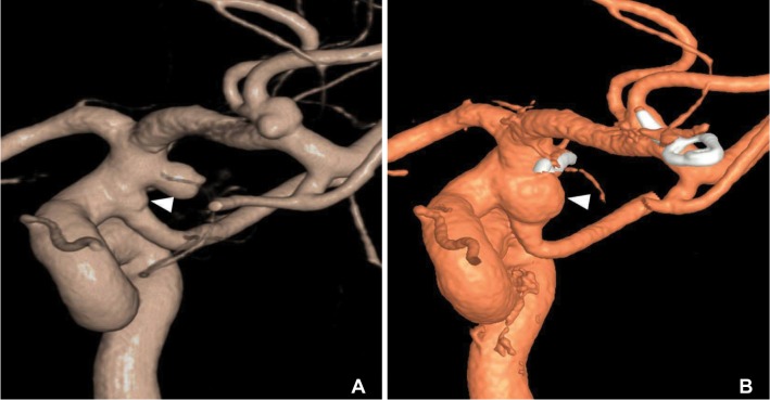
Fig. 2 A 57-year-old female (Case 2). (A) Initial angiogram showed a tiny bleb (arrowhead) at triangular junctional dilatation of the PcomA. (B) Follow-up angiogram obtained five years later revealed a de novo aneurysm formation from a junctional dilatation at the PcomA. It located just below the clipping site of the anterior choroidal artery aneurysm.
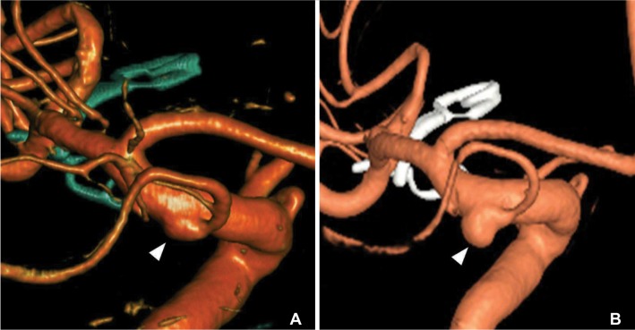
Fig. 3 A 55-year-old female (Case 3). (A) Initial angiogram showed a bleb (arrowhead) at junctional dilatation of the PcomA. (B) Follow-up angiogram obtained five years later revealed a de novo aneurysm (arrowhead) at the site of previous bleb.
Reference
-
1. Hassler O, Saltzman GF. Histologic changes in infundibular widening of the posterior communicating artery. A preliminary report. Acta Pathol Microbiol Scand. 1959; 46:305–312. PMID: 13852000.2. Taveras JM, Wood EH. Diagnostic Neuroradiology. Baltimore: Williams and Wilkins;1964. p. 498.3. Endo S, Furuichi S, Takaba M, Hirashima Y, Nishijima M, Takaku A. Clinical study of enlarged infundibular dilation of the origin of the posterior communicating artery. J Neurosurg. 1995; 83:421–425. PMID: 7666216.
Article4. Epstein F, Ransohoff J, Budzilovich GN. The clinical significance of junctional dilatation of the posterior communicating artery. J Neurosurg. 1970; 33:529–531. PMID: 5479491.
Article5. Itakura T, Ozaki F, Nakai E, Fujii T, Hayashi S, Komai N. Bilateral aneurysm formation developing from junctional dilatation (infundibulum) of the posterior communicating arteries. Case report. J Neurosurg. 1983; 58:117–119. PMID: 6847897.6. Fischer S, Hopf N, Henkes H. Evolution from an infundibulum of the posterior communicating artery to a saccular aneurysm. Clin Neuroradiol. 2011; 21:87–90.
Article7. Drake CG. On the surgical treatment of ruptured intracranial aneurysms. Clin Neurosurg. 1965; 13:122–155. PMID: 5870662.8. Stuntz JT, Ojemann GA, Alvord EC Jr. Radiographic and histologic demonstration of an aneurysm developing on the infundibulum of the posterior communicating artery. Case report. J Neurosurg. 1970; 33:591–595. PMID: 5479497.9. Trasi S, Vincent LM, Zingesser LH. Development of aneurysm from infundibulum of posterior communicating artery with documentation of prior hemorrhage. AJNR Am J Neuroradiol. 1981; 2:368–370. PMID: 6787905.10. Waga S, Morikawa A. Aneurysm developing on the infundibular widening of the posterior communicating artery. Surg Neurol. 1979; 11:125–127. PMID: 424981.11. Wollschlaeger G, Wollschlaeger PB, Lucas FV, Lopez VF. Experience and result with postmortem cerebral angiography performed as routine procedure of the autopsy. Am J Roentgenol Radium Ther Nucl Med. 1967; 101:68–87.
Article12. Yoshimoto T, Suzuki J. Surgical treatment of an aneurysm on the funnel-shaped bulge of the posterior communicating artery. Case report. J Neurosurg. 1974; 41:377–379. PMID: 4415999.13. Young B, Meacham WF, Allen JH. Documented enlargement and rupture of a small arterial sacculation. Case report. J Neurosurg. 1971; 34:814–817. PMID: 5561026.14. Coupe NJ, Athwal RK, Marshman LA, Brydon HL. Subarachnoid hemorrhage emanating from a ruptured infundibulum: case report and literature review. Surg Neurol. 2007; 67:204–206. PMID: 17254894.15. Marshman LA, Ward PJ, Walter PH, Dossetor RS. The progression of an infundibulum to aneurysm formation and rupture: case report and literature review. Neurosurgery. 1998; 43:1445–1448. discussion 1448-1449PMID: 9848859.
Article16. Radulovic D, Nestorovic B, Rakic M, Janosevic V. Enlargement to a saccular aneurysm and subsequent rupture of infundibular widening of posterior communicating artery. Neurochirurgie. 2006; 52:525–528. PMID: 17203900.
Article17. Takahashi C, Fukuda O, Hori E, Kameda H, Endo S. A case of infundibular dilatation developed into an aneurysm and rupturing after the rupture of an aneurysm 10 years ago. No Shinkei Geka. 2006; 34:613–617. PMID: 16768138.
- Full Text Links
-
- Actions
-
Cited
- CITED
-
- Close
- Share
-
- Similar articles
-
- Rupture of De Novo Anterior Communicating Artery Aneurysm 8 Days after the Clipping of Ruptured Middle Cerebral Artery Aneurysm
- Endovascular Embolization of a de Novo True Posterior Communicating Artery Aneurysm 23 years After Surgical Clipping of an Ipsilateral Posterior Communicating Artery-internal Carotid Artery Aneurysm: A Case Report
- Transposition of Anterior Choroidal Artery and Posterior Communicating Artery Origin
- An Aneurysm Developing on the Infundibulum of Posterior Communicating Artery: Case Report and Literature Review
- True Posterior Communicating Artery Aneurysm
