Korean J Urol.
2015 May;56(5):337-345. 10.4111/kju.2015.56.5.337.
Current role of multiparametric magnetic resonance imaging in the management of prostate cancer
- Affiliations
-
- 1Department of Surgery, Austin Hospital, University of Melbourne, Melbourne, VIC, Australia. lawrentschuk@gmail.com
- 2Sydney Adventist Hospital, Sydney, NSW, Australia.
- 3Olivia Newton-John Cancer Research Institute, Austin Hospital, Heidelberg, VIC, Australia.
- 4Division of Cancer Surgery, Peter MacCallum Cancer Centre, Melbourne, VIC, Australia.
- KMID: 2155312
- DOI: http://doi.org/10.4111/kju.2015.56.5.337
Abstract
- The purpose of this review was to evaluate the current role of multiparametric magnetic resonance imaging (mp-MRI) in the management of prostate cancer (PC). The diagnosis of PC remains controversial owing to overdetection of indolent disease, which leads to overtreatment and subsequent patient harm. mp-MRI has the potential to equilibrate the imbalance between detection and treatment. The limitation of the data for analysis with this new technology is problematic, however. This issue has been compounded by a paradigm shift in clinical practice aimed at utilizing this modality, which has been rolled out in an ad hoc fashion often with commercial motivation. Despite a growing body of literature, pertinent clinical questions remain. For example, can mp-MRI be calibrated to reliably detect biologically significant disease? As with any new technology, objective evaluation of the clinical applications of mp-MRI is essential. The focus of this review was on the evaluation of mp-MRI of the prostate with respect to clinical utility.
MeSH Terms
Figure
-

Fig. 1 T2-weighted image demonstrating an area of low signal intensity in the right peripheral zone consistent with prostate cancer.

Fig. 2 A dynamic contrast enhancement image (A) and postcontrast washout curve (B) of a right peripheral zone lesion.
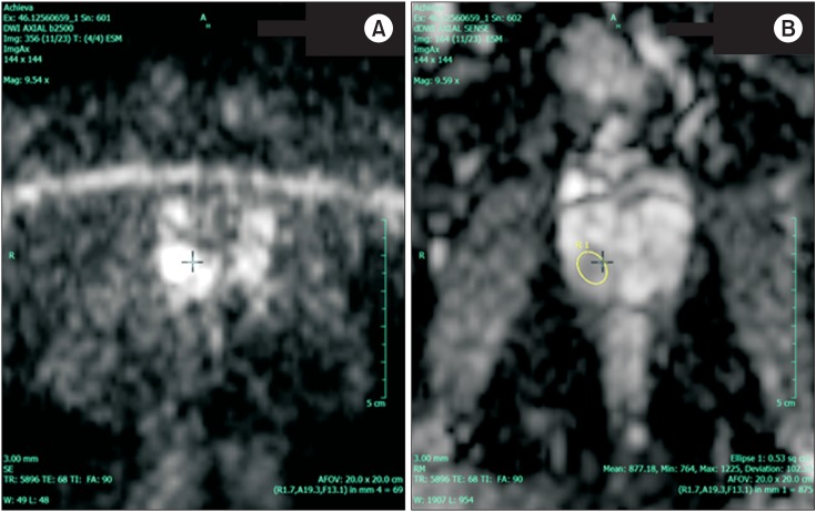
Fig. 3 A diffusion-weighted image (b value= 2,500) (A) and the corresponding apparent diffusion coefficient map (B) demonstrating Gleason 4+3 prostate cancer in the right peripheral zone.
Reference
-
1. Schroder FH. Screening for prostate cancer: current status of ERSPC and screening-related issues. Recent Results Cancer Res. 2014; 202:47–51. PMID: 24531776.2. Moyer VA. U.S. Preventive Services Task Force. Screening for prostate cancer: U.S. Preventive Services Task Force recommendation statement. Ann Intern Med. 2012; 157:120–134. PMID: 22801674.
Article3. Phillips R. Bladder cancer: Stem cells repopulate tumours. Nat Rev Urol. 2015; 12:63. PMID: 25535001.4. Dall'Era MA, Albertsen PC, Bangma C, Carroll PR, Carter HB, Cooperberg MR, et al. Active surveillance for prostate cancer: a systematic review of the literature. Eur Urol. 2012; 62:976–983. PMID: 22698574.5. Wilt TJ, Brawer MK, Jones KM, Barry MJ, Aronson WJ, Fox S, et al. Radical prostatectomy versus observation for localized prostate cancer. N Engl J Med. 2012; 367:203–213. PMID: 22808955.
Article6. Pokorny MR, de Rooij M, Duncan E, Schroder FH, Parkinson R, Barentsz JO, et al. Prospective study of diagnostic accuracy comparing prostate cancer detection by transrectal ultrasoundguided biopsy versus magnetic resonance (MR) imaging with subsequent MR-guided biopsy in men without previous prostate biopsies. Eur Urol. 2014; 66:22–29. PMID: 24666839.
Article7. Thompson J, Lawrentschuk N, Frydenberg M, Thompson L, Stricker P. USANZ. The role of magnetic resonance imaging in the diagnosis and management of prostate cancer. BJU Int. 2013; 112(Suppl 2):6–20. PMID: 24127671.
Article8. Raz O, Haider M, Trachtenberg J, Leibovici D, Lawrentschuk N. MRI for men undergoing active surveillance or with rising PSA and negative biopsies. Nat Rev Urol. 2010; 7:543–551. PMID: 20930867.
Article9. Panebianco V, Barchetti F, Sciarra A, Ciardi A, Indino EL, Papalia R, et al. Multiparametric magnetic resonance imaging vs. standard care in men being evaluated for prostate cancer: a randomized study. Urol Oncol. 2015; 33:17e1–e7. PMID: 25443268.
Article10. Barentsz JO, Richenberg J, Clements R, Choyke P, Verma S, Villeirs G, et al. ESUR prostate MR guidelines 2012. Eur Radiol. 2012; 22:746–757. PMID: 22322308.
Article11. Roy C, Foudi F, Charton J, Jung M, Lang H, Saussine C, et al. Comparative sensitivities of functional MRI sequences in detection of local recurrence of prostate carcinoma after radical prostatectomy or external-beam radiotherapy. AJR Am J Roentgenol. 2013; 200:W361–W368. PMID: 23521479.
Article12. Yacoub JH, Oto A, Miller FH. MR imaging of the prostate. Radiol Clin North Am. 2014; 52:811–837. PMID: 24889173.
Article13. Katahira K, Takahara T, Kwee TC, Oda S, Suzuki Y, Morishita S, et al. Ultra-high-b-value diffusion-weighted MR imaging for the detection of prostate cancer: evaluation in 201 cases with histopathological correlation. Eur Radiol. 2011; 21:188–196. PMID: 20640899.
Article14. Luczynska E, Heinze-Paluchowska S, Domalik A, Cwierz A, Kasperkiewicz H, Blecharz P, et al. The utility of diffusion weighted imaging (DWI) using apparent diffusion coefficient (ADC) values in discriminating between prostate cancer and normal tissue. Pol J Radiol. 2014; 79:450–455. PMID: 25484999.
Article15. Donati OF, Afaq A, Vargas HA, Mazaheri Y, Zheng J, Moskowitz CS, et al. Prostate MRI: evaluating tumor volume and apparent diffusion coefficient as surrogate biomarkers for predicting tumor Gleason score. Clin Cancer Res. 2014; 20:3705–3711. PMID: 24850842.
Article16. Boesen L, Chabanova E, Logager V, Balslev I, Thomsen HS. Apparent diffusion coefficient ratio correlates significantly with prostate cancer gleason score at final pathology. J Magn Reson Imaging. 2014; 11. 19. [Epub]. http://dx.doi.org/10.1002/jmri.24801.
Article17. Weinreb JC, Blume JD, Coakley FV, Wheeler TM, Cormack JB, Sotto CK, et al. Prostate cancer: sextant localization at MR imaging and MR spectroscopic imaging before prostatectomy--results of ACRIN prospective multi-institutional clinicopathologic study. Radiology. 2009; 251:122–133. PMID: 19332850.
Article18. Bratan F, Niaf E, Melodelima C, Chesnais AL, Souchon R, Mege-Lechevallier F, et al. Influence of imaging and histological factors on prostate cancer detection and localisation on multiparametric MRI: a prospective study. Eur Radiol. 2013; 23:2019–2029. PMID: 23494494.
Article19. Rosenkrantz AB, Kim S, Lim RP, Hindman N, Deng FM, Babb JS, et al. Prostate cancer localization using multiparametric MR imaging: comparison of Prostate Imaging Reporting and Data System (PI-RADS) and Likert scales. Radiology. 2013; 269:482–492. PMID: 23788719.
Article20. Portalez D, Mozer P, Cornud F, Renard-Penna R, Misrai V, Thoulouzan M, et al. Validation of the European Society of Urogenital Radiology scoring system for prostate cancer diagnosis on multiparametric magnetic resonance imaging in a cohort of repeat biopsy patients. Eur Urol. 2012; 62:986–996. PMID: 22819387.
Article21. Vache T, Bratan F, Mege-Lechevallier F, Roche S, Rabilloud M, Rouviere O. Characterization of prostate lesions as benign or malignant at multiparametric MR imaging: comparison of three scoring systems in patients treated with radical prostatectomy. Radiology. 2014; 272:446–455. PMID: 24937690.
Article22. Junker D, Quentin M, Nagele U, Edlinger M, Richenberg J, Schaefer G, et al. Evaluation of the PI-RADS scoring system for mpMRI of the prostate: a whole-mount step-section analysis. World J Urol. 2014; 8. 1. [Epub]. http://dx.doi.org/10.1007/s00345-014-1370-x.
Article23. Styles C, Ferris N, Mitchell C, Murphy D, Frydenberg M, Mills J, et al. Multiparametric 3T MRI in the evaluation of intraglandular prostate cancer: correlation with histopathology. J Med Imaging Radiat Oncol. 2014; 58:439–448. PMID: 24935089.
Article24. Kitamura K, Muto S, Yokota I, Hoshimoto K, Kaminaga T, Noguchi T, et al. Feasibility of multiparametric prostate magnetic resonance imaging in the detection of cancer distribution: histopathological correlation with prostatectomy specimens. Prostate Int. 2014; 2:188–195. PMID: 25599075.
Article25. Billing A, Buchner A, Stief C, Roosen A. Preoperative mp-MRI of the prostate provides little information about staging of prostate carcinoma in daily clinical practice. World J Urol. 2014; 11. 29. [Epub]. http://dx.doi.org/10.1007/s00345-014-1448-5.
Article26. Min BD, Kim WT, Cho BS, Kim YJ, Yun SJ, Lee SC, et al. Usefulness of a combined approach of t1-weighted, t2-weighted, dynamic contrast-enhanced, and diffusion-weighted imaging in prostate cancer. Korean J Urol. 2012; 53:830–835. PMID: 23301126.
Article27. Tanaka K, Shigemura K, Muramaki M, Takahashi S, Miyake H, Fujisawa M. Efficacy of using three-tesla magnetic resonance imaging diagnosis of capsule invasion for decision-making about neurovascular bundle preservation in robotic-assisted radical prostatectomy. Korean J Urol. 2013; 54:437–441. PMID: 23878685.
Article28. Le JD, Huang J, Marks LS. Targeted prostate biopsy: value of multiparametric magnetic resonance imaging in detection of localized cancer. Asian J Androl. 2014; 16:522–529. PMID: 24589455.
Article29. Pepe P, Garufi A, Priolo G, Pennisi M. Can 3-Tesla pelvic phased-array multiparametric MRI avoid unnecessary repeat prostate biopsy in patients with PSA < 10 ng/mL? Clin Genitourin Cancer. 2015; 13:e27–e30. PMID: 25081324.30. Le JD, Stephenson S, Brugger M, Lu DY, Lieu P, Sonn GA, et al. Magnetic resonance imaging-ultrasound fusion biopsy for prediction of final prostate pathology. J Urol. 2014; 192:1367–1373. PMID: 24793118.
Article31. Afshar-Oromieh A, Malcher A, Eder M, Eisenhut M, Linhart HG, Hadaschik BA, et al. PET imaging with a [68Ga]galliumlabelled PSMA ligand for the diagnosis of prostate cancer: biodistribution in humans and first evaluation of tumour lesions. Eur J Nucl Med Mol Imaging. 2013; 40:486–495. PMID: 23179945.
Article32. Afshar-Oromieh A, Avtzi E, Giesel FL, Holland-Letz T, Linhart HG, Eder M, et al. The diagnostic value of PET/CT imaging with the (68)Ga-labelled PSMA ligand HBED-CC in the diagnosis of recurrent prostate cancer. Eur J Nucl Med Mol Imaging. 2015; 42:197–209. PMID: 25411132.
Article33. Schoots IG, Petrides N, Giganti F, Bokhorst LP, Rannikko A, Klotz L, et al. Magnetic Resonance Imaging in Active Surveillance of Prostate Cancer: A Systematic Review. Eur Urol. 2015; 67:627–636. PMID: 25511988.
Article34. Abdi H, Pourmalek F, Zargar H, Walshe T, Harris AC, Chang SD, et al. Multiparametric magnetic resonance imaging enhances detection of significant tumor in patients on active surveillance for prostate cancer. Urology. 2015; 85:423–428. PMID: 25623709.
Article35. WaltonDiaz A, Shakir NA, George AK, Rais-Bahrami S, Turkbey B, Rothwax JT, et al. Use of serial multiparametric magnetic resonance imaging in the management of patients with prostate cancer on active surveillance. Urol Oncol. 2015; 3. 05. [Epub]. http://dx.doi.org/10.1016/j.urolonc.2015.01.023.36. Siddiqui MM, Truong H, Rais-Bahrami S, Stamatakis L, Logan J, Walton-Diaz A, et al. Clinical Implications of a Multiparametric Magnetic Resonance Imaging Based Nomogram Applied to Prostate Cancer Active Surveillance. J Urol. 2015; 1. 26. [Epub]. http://dx.doi.org/10.1016/j.juro.2015.01.088.
Article37. Ha JY, Kim BH, Park CH, Kim CI. Early experience with active surveillance in low-risk prostate cancer treated. Korean J Urol. 2014; 55:167–171. PMID: 24648870.
Article
- Full Text Links
-
- Actions
-
Cited
- CITED
-
- Close
- Share
-
- Similar articles
-
- Multiparametric MRI in the Detection of Clinically Significant Prostate Cancer
- Multiparametric magnetic resonance imaging for prostate cancer: A review and update for urologists
- The Use of Magnetic Resonance Imaging in the Prostate Cancer Primary Diagnostic Pathway: Is It Ready for Primetime?
- Prostate Imaging Reporting and Data System (PI-RADS) v 2.1: Overview and Critical Points
- Are we ready to adopt the European Association of Urology recommendations on multiparametric magnetic resonance imaging in the early detection of prostate cancer?
